
A rapid segmentation method of aortic valve based on esophageal ultrasound
A technique of aortic valve and ultrasound, applied in image analysis, image data processing, instruments, etc., can solve problems such as incomplete segmentation and overflow of ultrasound images
- Summary
- Abstract
- Description
- Claims
- Application Information
AI Technical Summary
Problems solved by technology
Method used
Image

Examples
Embodiment
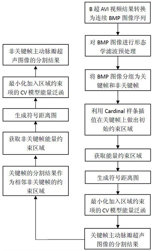
[0046] The invention is Dual-CoreCPUE58003.20GHz, the graphics card is NVIDIAGeForceGT430NVIDIAGeForceGT430, the memory is 2.00GB, the operating system is WindowXP computer, and the whole segmentation method is written in C++ and Matlab language.
[0047] (1) The B-ultrasound video results (AVI format) output by transesophageal ultrasound use the DirectShow platform and the FFDShow video format decoder to convert the AVI format video files into 24-bit or 8-bit BMP format continuous ultrasound image sequences. Arrange the ultrasound image sequence in chronological order.
[0048] (2) Perform morphological filtering preprocessing on the above continuous image sequence: perform closed operation on the original image of the ultrasonic image to obtain the marked image, then perform erosion operation on the marked image and perform intersection operation with the original image until the iteration ends when convergence. The preprocessed ultrasound image can not only reduce the spe...
PUM
 Abstract
Description
Claims
Application Information