3D hysteroscope surgery device with visible whole course

A surgical device and a hysteroscope technology, applied in the field of medical devices, can solve the problems of unclear display of protruding parts in the patient's body, inability to accurately control the flushing part, and unsatisfactory display accuracy, and achieve compact structure and increased momentum. , the effect of small outer diameter

Active Publication Date: 2020-07-31
THE SECOND AFFILIATED HOSPITAL OF CHONGQING MEDICAL UNIV
View PDF12 Cites 0 Cited by
  • Summary
  • Abstract
  • Description
  • Claims
  • Application Information

AI Technical Summary

Problems solved by technology

[0003] In the existing hysteroscopic surgery, a large tube and a small tube are usually used, the small tube is set in the large tube, the tube hole of the small tube is used as the operation hole, and the operation hole is used to extend into the surgical instrument, and the gap between the small tube and the large tube is used to Flush water (or other liquid) to the surgical site to flush blood or tissue debris, etc., which is convenient for the operator to observe, but since the water inlet and drainage are located in the same channel, the channel is small and only a small amount of water can be flushed, and a large amount of flushing is required When watering, it is often necessary to fill the uterus before releasing the water. The process is too cumbersome, wastes time, increases the risk of surgery, and the flushing position cannot be accurately controlled to ensure the effectiveness of flushing.
In addition, the image provided by hysteroscopic surgery is a flat image, which is different from the actual effect of human eyes. The prominent parts in the patient's body are not clearly displayed, and the accuracy of the display is not ideal.

Method used


Image

  • 3D hysteroscope surgery device with visible whole course
  • 3D hysteroscope surgery device with visible whole course
  • 3D hysteroscope surgery device with visible whole course

Examples


Embodiment Construction

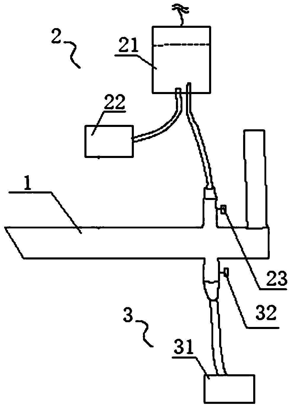
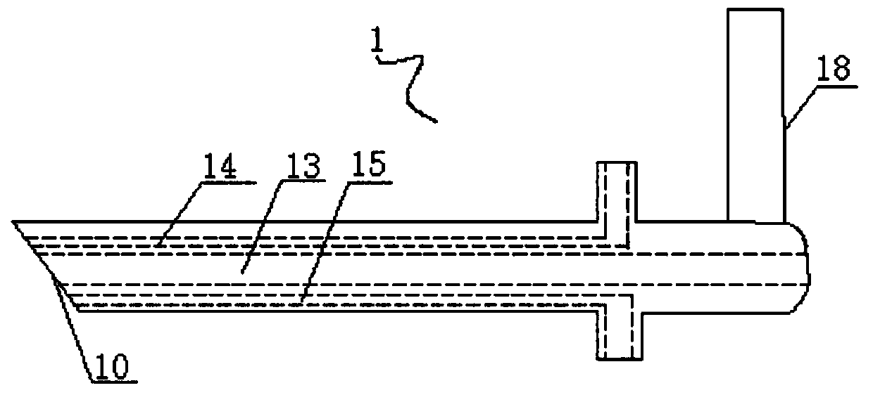
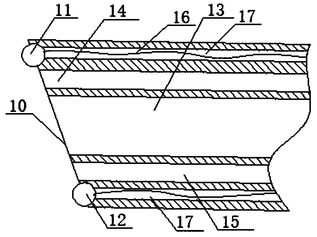
[0045] Embodiments of the present invention are described in detail below, examples of which are shown in the drawings, wherein the same or similar reference numerals designate the same or similar elements or elements having the same or similar functions throughout. The embodiments described below by referring to the figures are exemplary only for explaining the present invention and should not be construed as limiting the present invention.

[0046] In describing the present invention, it should be understood that the terms "longitudinal", "transverse", "upper", "lower", "front", "rear", "left", "right", "vertical", The orientation or positional relationship indicated by "horizontal", "top", "bottom", "inner", "outer", etc. are based on the orientation or positional relationship shown in the drawings, and are only for the convenience of describing the present invention and simplifying the description, rather than Nothing indicating or implying that a referenced device or elem...


PUM


Abstract

The invention discloses a 3D hysteroscope surgery device capable of being seen in the whole process. The device comprises a mirror rod, infusion equipment, waste liquid collecting equipment, a surgical instrument, processing equipment and display equipment; an operation hole, at least one first flow channel for spraying liquid outwards and at least one second flow channel for recovering liquid areformed in the mirror rod; the front end face of the mirror rod is inclined, and a liquid outlet of the first flow channel is located at the far end of the front end face; a first image sensor and a second image sensor are arranged on the front end surface; a liquid outlet of the infusion equipment is connected to a liquid inlet of the mirror rod, and a liquid inlet of the waste liquid collectingequipment is connected to a liquid outlet of the mirror rod; and the processing equipment receives and processes an image signal, and outputs a display signal to the display equipment to display a stereoscopic image. A surgical site is displayed in a three-dimensional mode, so that the protruding part is displayed more clearly, the mirror rod improves the flushing effectiveness, a liquid circulation loop is formed, a large amount of continuous flushing can be achieved, flushing operation is simplified, and flushing is more convenient and faster.

Description

technical field [0001] The invention belongs to the technical field of medical devices, in particular to a whole-process visual 3D hysteroscopic operation device. Background technique [0002] In the case of uterine fibroids, uterine cancer, or unwanted pregnancy, it is necessary to perform surgery in the uterus. With the need for visualization of surgery, it is becoming more and more popular and common to use hysteroscope with surgical instruments for surgery. For example, the existing abortion is mainly divided into negative pressure suction, curettage, hysteroscopic abortion and so on. Negative pressure aspiration is suitable for the uterus within 10 weeks of pregnancy. It mainly uses the method of negative pressure electric aspiration to suck out the early pregnancy products, namely the embryo sac and decidua tissue. The curettage range of forceps curettage is generally advocated within 14 weeks. For those who require termination of pregnancy or are not suitable for pre...

Claims


Application Information

Patent Timeline
Patent Type & Authority Applications(China)
IPC IPC(8): A61B90/00A61B18/12
CPCA61B90/37A61B90/361A61B18/12A61B2090/371A61B2217/005A61B2217/007A61B17/42A61B17/1204A61B2018/00559A61B2018/00589A61B2218/007A61B2218/002Y02P70/10
Inventor 李晓霞郎中兵杨杰
Owner THE SECOND AFFILIATED HOSPITAL OF CHONGQING MEDICAL UNIV
Why Eureka
Try Eureka