Scanning method and device for multi-modal medical equipment

A technology of medical equipment and scanning methods, applied in the medical field, can solve problems such as low efficiency and high labor costs

Pending Publication Date: 2020-11-10
SHANGHAI UNITED IMAGING HEALTHCARE
View PDF3 Cites 5 Cited by
  • Summary
  • Abstract
  • Description
  • Claims
  • Application Information

AI Technical Summary

Problems solved by technology

[0004] The present application provides a scanning method and device for multimodal medical equipment, so as to at least solve the problem of high labor costs and low efficiency in the process of finding or confirming abnormal points in the related art

Method used


Image

  • Scanning method and device for multi-modal medical equipment
  • Scanning method and device for multi-modal medical equipment
  • Scanning method and device for multi-modal medical equipment

Examples


Embodiment Construction

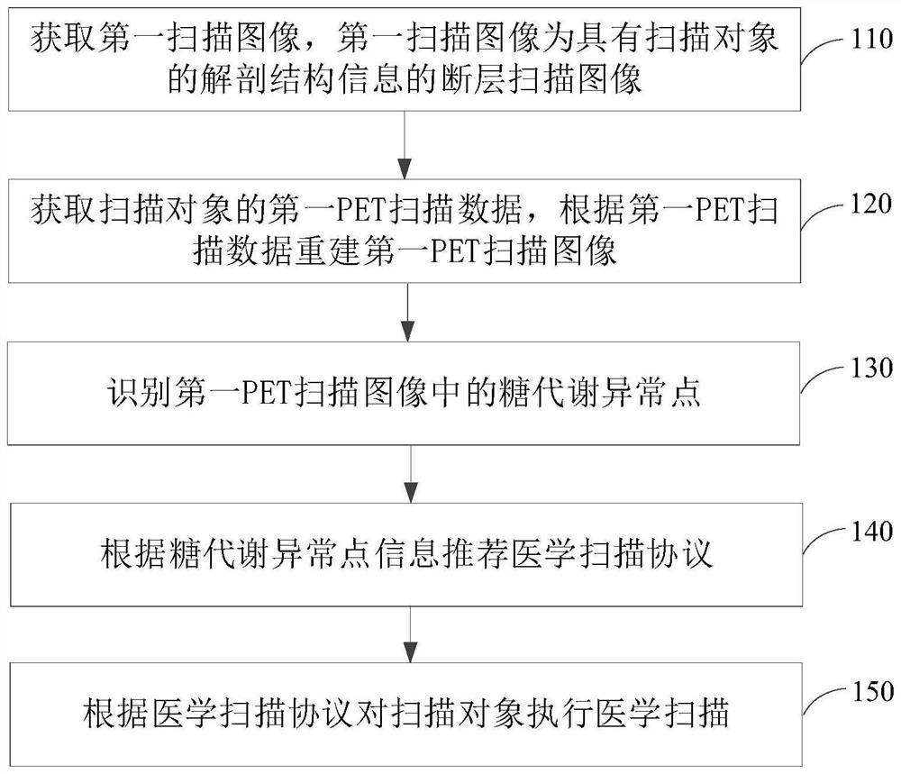
[0045] In order to make the purpose, technical solutions and advantages of the present application clearer, the present application will be described and illustrated below in conjunction with the accompanying drawings and embodiments. It should be understood that the specific embodiments described here are only used to explain the present application, and are not intended to limit the present application. Based on the embodiments provided in the present application, all other embodiments obtained by persons of ordinary skill in the art without creative efforts shall fall within the protection scope of the present application.

[0046] Obviously, the accompanying drawings in the following description are only some examples or embodiments of the present application, and those skilled in the art can also apply the present application to other similar scenarios. In addition, it can also be understood that although such development efforts may be complex and lengthy, for those of ...


PUM


Abstract

The invention relates to a scanning method and device for multi-modal medical equipment. The method comprises the steps of obtaining a first scanning image which is a tomography image with the anatomical structure information of a scanned object; acquiring the first PET scanning data of the scanned object, and reconstructing a first PET scanning image according to the first PET scanning data; identifying an abnormal glucose metabolism point in the first PET scanning image; recommending a medical scanning protocol according to the abnormal glucose metabolism point information; and performing medical scanning on the scanned object according to the medical scanning protocol. According to the method, the abnormal point in the PET scanning image is automatically identified, and the medical scanning protocol is intelligently recommended based on the position with glucose metabolism abnormality, so that the problems of relatively higher labor cost and relatively lower efficiency in an abnormal point discovery or confirmation process in related technologies are solved.

Description

technical field [0001] The present application relates to the field of medical technology, in particular to a scanning method and device for multimodal medical equipment. Background technique [0002] Existing medical imaging examinations include methods such as CT, DR, MRI, PET-CT, and PETMR. These examinations all need to obtain scanned images by selecting one or more protocol groups. During the scan execution, after obtaining partial scan results, the user usually scans some other types of protocols due to clinical diagnosis. For example, after a sagittal or coronal scan of the spine, some abnormal lesions are found. If you want to take a closer look at some information about the spine, and want to perform a more detailed transverse scan of the intervertebral disc or vertebral body, you need Select the scan protocol group corresponding to the traversal bit or add the protocol to the list for scanning. As another example, in the PET scanning process, after the PET image ...

Claims


Application Information

Patent Timeline
Patent Type & Authority Applications(China)
IPC IPC(8): A61B5/00A61B6/00
CPCA61B5/0037A61B5/0033A61B6/488
Inventor 顾笑悦李金龙贾胜果
Owner SHANGHAI UNITED IMAGING HEALTHCARE
Why Eureka
Try Eureka