Preparation method and application of magnetic control fluorescent biosensor

A biosensor and fluorescence technology, applied in the field of biosensors, can solve the problems of large size, inability to meet rapid detection, poor specificity, etc.

Pending Publication Date: 2021-06-11
GUANGXI NORMAL UNIV
View PDF6 Cites 2 Cited by
  • Summary
  • Abstract
  • Description
  • Claims
  • Application Information

AI Technical Summary

Problems solved by technology

[0003] Among the relevant patents for the detection of tumor markers that have been published so far, CN109900911A is a method for detecting liver cancer tumor marker AFP by using core-shell nanostars, using the core-shell nanostars as the substrate, and using the principle of surface-enhanced Raman scattering to detect , although the method has good sensitivity and specificity, it needs multiple modification and functionalization of the substrate, and the method is not simple enough
[0004] CN106807308A A mass spectrometry method for extracting tumor markers in serum using magnetic nanoparticles with a core-shell structure. The method utilizes magnetic core-shell materials to enrich and separate tumor markers from serum, and quantifies them with mass spectrometry. Although it can be quickly Efficient detection, but this method has poor specificity, easily leads to false positive results, and requires the use of large instruments and sample pretreatment. The operation process is cumbersome and complicated, and the instruments are expensive and bulky, which cannot meet the needs of on-site rapid detection
[0005] In addition, among the existing sensing analysis methods based on signal amplification for sensitive detection of tumor markers, CN111351934A is a method for electrochemical detection of lung cancer tumor markers by co-modifying electrodes with hollow gold nanospheres and nano-petal-shaped manganese dioxide , the synerg

Method used


Image

  • Preparation method and application of magnetic control fluorescent biosensor
  • Preparation method and application of magnetic control fluorescent biosensor

Examples


Embodiment

[0043] A method for preparing a magnetically controlled fluorescent biosensor based on multiple signal amplification, comprising the following steps:

[0044] (1) Preparation of MB-Ab magnetically controlled immune probe;

[0045] Take 1mL MB (5mg mL -1 ) for magnetic separation and washed 2-3 times with 1 mL of PBS (0.01 M, pH=7.4);

[0046] After washing, add 1 mL of EDC (0.0766 g) and shake at room temperature for 10 min, then add 0.0114 g of NHS and continue shaking for 30 min, magnetically separate and wash with PBS 2-3 times;

[0047] Then add 1 mL of PBS (0.01M) and 100 μL of anti-CEA antibody, shake at room temperature for 3 h, then magnetically separate and wash with PBS for 2-3 times, the above obtained substances are dispersed in 1 mL of 2.0 wt% BSA and shaken for 1 h, and finally stored in a refrigerator at 4°C until use.

[0048] (2) Preparation of liposome-embedded gold cluster complex Biotin-Liposome / AuNCs;

[0049] Accurately weigh 75 mg of BSA into a 100 m...

experiment example

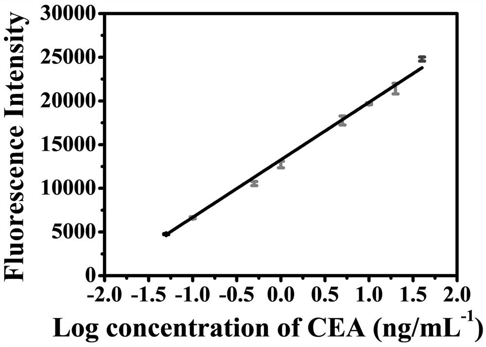
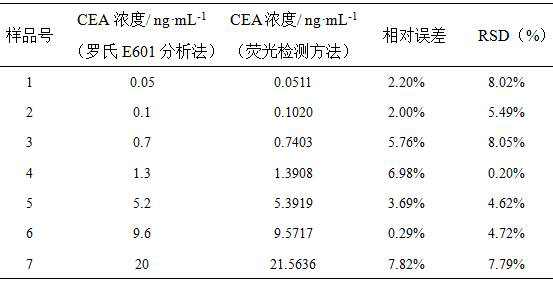
[0062] Serum samples were tested: 5 clinical serum samples were retrieved from Guilin Hospital of Integrated Traditional Chinese and Western Medicine, and the concentrations of CEA in the serum samples were 0.7, 1.3, 5.2, 9.6, and 20 ng / mL, respectively. Take 10 μL of serum samples of 20 ng / mL CEA and add them to two 5 mL centrifuge tubes, then add PBS (10 mM, pH 7.4) solution to make the final concentration of CEA in the samples 0.05 ng / mL and 0.1 ng / mL, Serum samples containing CEA concentrations of 0.05, 0.1, 0.7, 1.3, 5.2, 9.6, and 20 ng / mL were numbered 1, 2, 3, 4, 5, 6, and 7, respectively. Next, the above 7 samples were detected by magnetically controlled fluorescent immunoassay, and the signal values ​​generated by the 7 samples were recorded, and substituted into the working curve to obtain the concentration of CEA in the serum sample, as shown in Table 1:

[0063] Table 1 Comparison of experimental results of CEA antigen in human serum measured by magnetically contro...


PUM


Abstract

The invention discloses a preparation method and application of a magnetic control fluorescent biosensor. The preparation method comprises the following steps of (1) preparing a magnetic control immune probe MB-Ab; (2) preparing a liposome embedded gold cluster compound Biotin-Liposome/AuNCs (gold nano-particles); (3) preparing an aptamer modified lipidosome embedded gold cluster compound, namely, a Biotin-Liposome/AuNCs-Aptamer signal probe, (4) a Biotin-Liposome/AuNCs-Aptamer signal probe being combined with a magnetic control immune probe to construct a multiple signal amplification magnetic control fluorescence biosensor for detecting the carcino-embryonic antigen (CEA); during detection, a fluorescence signal working curve of the sensor for detecting the carcino-embryonic antigens with different concentrations being drawn firstly, and then the concentration of the carcino-embryonic antigens in a clinical serum sample being detected. The method is advantaged in that the magnetic control fluorescent biosensor prepared by the preparation method disclosed by the invention is strong in anti-interference capability, has high sensitivity and high selectivity, and can be used for detecting low-abundance tumor markers in a serum sample.

Description

technical field [0001] The invention relates to a biosensor, in particular to a preparation method and application of a magnetically controlled fluorescent biosensor based on multiple signal amplification. Background technique [0002] Tumor markers, also known as tumor markers, are substances synthesized, released, and secreted by tumor cells during the process of tumorigenesis and proliferation, or produced by a host's response to tumor cells. The analysis of the content of tumor markers can be used as an assistant to judge whether there is a tumor clinically, and it is very helpful for the early detection of some cancers. In addition, the detection of tumor markers can also monitor the development of tumor screening and the efficacy after treatment. Enzyme-linked immunosorbent assay (ELISA) has become a main method for detecting tumor markers due to its simple operation and rapidity, but the detection of low-abundance tumor markers often requires a signal amplification s...

Claims


Application Information

Patent Timeline
IPC IPC(8): G01N33/574G01N33/535G01N21/64
CPCG01N33/57473G01N33/535G01N21/6402G01N21/6428G01N2021/6439Y02A50/30
Inventor 侯丽黄娟娟蒋高艳林天然赵书林
Owner GUANGXI NORMAL UNIV
Why Eureka
Try Eureka