Supercharge Your Innovation With Domain-Expert AI Agents!

Image diagnosis support apparatus, medical image acquisition apparatus, and computer readable recording medium

A medical image and image diagnosis technology, applied in the direction of diagnostic recording/measurement, computer-aided medical procedures, diagnosis, etc., can solve problems such as artifacts, undiagnosable, unsolvable, etc.

Active Publication Date: 2021-07-09
HITACHI HEALTHCARE MFG LTD
View PDF13 Cites 0 Cited by
  • Summary
  • Abstract
  • Description
  • Claims
  • Application Information

AI Technical Summary

Problems solved by technology

[0006] In image diagnosis, when measuring multiple images of the same subject and selecting and using one or more images that are most suitable for the object of diagnosis from these multiple images, due to the influence of body movement, etc. or according to shooting conditions etc., artifacts may occur in parts or areas that should be diagnosed in the image that should be selected, making it impossible to use for diagnosis
In this case, although other images are considered as alternatives, their selection is not easy
This problem also exists even for images obtained in different modalities of image types, such as MR images, CT images, X-ray images, etc., and the technology described in Patent Document 1 cannot solve it
[0007] In addition, even if a predetermined image is selected, the contrast of the image will vary depending on the manufacturer (supplier) of the medical image acquisition device and the imaging conditions. For example, in the case of an MRI apparatus, the contrast of the image will vary depending on the magnetic field strength and imaging parameters. , the selected image may not have the same contrast as the image used to generate the learned AI, so when using the existing AI, it is impossible to perform image diagnosis assistance with high accuracy

Method used


Image

  • Image diagnosis support apparatus, medical image acquisition apparatus, and computer readable recording medium
  • Image diagnosis support apparatus, medical image acquisition apparatus, and computer readable recording medium
  • Image diagnosis support apparatus, medical image acquisition apparatus, and computer readable recording medium

Examples


no. 1 approach >

[0042] In this embodiment, one image is selected from a plurality of images, and after the image is converted into an image for detection, the corresponding disease or lesion is detected and presented.

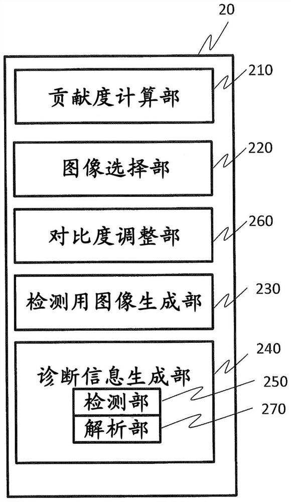
[0043] The configuration of the image processing unit 200 of the present embodiment and figure 2 The configuration shown is the same, and includes an image selection unit 220 that selects one of a plurality of images based on the contribution degree calculated by the contribution degree calculation unit 210 . In addition, the detection image generation unit 230 includes a contrast adjustment unit 260A. Figure 4 (A) and (B) in (A) show details of the contribution degree calculation unit 210 and the detected image generation unit 230 . The contribution calculation unit 210 includes: an artifact detection unit 211 that detects artifacts in an image; a reliability calculation unit 213 that digitizes the artifacts and calculates them as reliability; uses the reliability sum to d...

Deformed example 1

[0059] In the above-mentioned embodiments, the case of using the presence or absence of artifacts and the size of artifacts as indicators of the reliability of a plurality of images has been described, but instead of artifacts, or in addition to artifacts, the SNR of images may be used. Additionally the SNR of the image is used. The SNR of an image can be obtained by a known method such as a method of obtaining the SNR from the average value and standard deviation of the pixel values ​​of an arbitrarily set region of interest, and can be calculated by normalizing the SNR calculated for each image. reliability. In addition, the reliability calculated from artifacts and the reliability calculated from SNR may be weighted and summed as the reliability.

Deformed example 2

[0061] In the above-mentioned embodiment, the case where the contrast adjustment unit 260A adjusts the contrast of the selected image to the contrast of the learning image of the detection unit 250 has been described, but when the selected image is an MR image, images with different contrasts can also be adjusted For contrast adjustment between types, use another contrast image as a selection image. For example, when the image selected by the image selection unit 220 is a T1-weighted image, but the T2-weighted image is suitable for the diagnosis object, after adjusting the contrast of the T1-weighted image to match the contrast of the unused T2-weighted image, it is adjusted to Contrast of T2-weighted images as learning images. Although it is also possible to directly adjust the contrast of the learning image, by performing contrast adjustment on the measured T2-weighted image in this way, it is possible to perform contrast adjustment while maintaining the original information...


PUM


Abstract

The invention provides an image diagnosis support apparatus, a medical image acquisition apparatus, and a computer readable recording medium. The most appropriate image for a diagnostic target among a plurality of images is selected and accurate diagnosis support information is presented regardless of the type of a selected image, a modality, or the like. The image diagnosis support apparatus includes: a diagnostic information generation unit that generates diagnostic information based on a plurality of medical images; a reliability calculation unit that evaluates an image quality and calculates an image reliability for each of the plurality of medical images; and a degree-of-contribution calculation unit that calculates a degree of contribution of each of the plurality of medical images to the diagnostic information using an internal parameter indicating a degree of appropriateness of each medical image for a diagnostic target and the reliability calculated by the reliability calculation unit. An image for detection used by the diagnostic information generation unit is generated based on the degree of contribution.

Description

technical field [0001] The present invention relates to a technique for assisting diagnosis using images acquired by medical image acquisition devices such as magnetic resonance imaging devices (hereinafter referred to as MRI devices), X-ray imaging devices, and CT devices. Background technique [0002] In the medical field, image diagnosis for diagnosing diseases and lesions based on images captured by MRI apparatuses and CT apparatuses is widely performed. In recent years, a diagnostic support technology has also been developed that uses AI obtained by machine learning to determine the presence or absence of lesions, the degree of progression, or the degree of malignancy in measured images (Non-Patent Document 1, etc.). [0003] In support of image diagnosis using machine learning, AI is obtained by learning the relationship between them and images for each type of disease and each part, and one or more measurement images are used as input to determine the presence or abse...

Claims


Application Information

Patent Timeline
Patent Type & Authority Applications(China)
IPC IPC(8): G06T7/00G06T7/62G16H50/20A61B6/03
CPCG06T7/0012G06T7/62G16H50/20A61B6/03G06T2207/10088G16H30/40G06T2207/20084G06T2207/30016G06T2207/30096G01R33/5608G01R33/5602A61B6/5217A61B6/5235A61B6/5258G06N3/045G06T7/0014A61B5/055G06N20/00A61B5/0042
Inventor 佐藤良太雨宫知树白猪亨尾藤良孝
Owner HITACHI HEALTHCARE MFG LTD
Features
  • R&D
  • Intellectual Property
  • Life Sciences
  • Materials
  • Tech Scout
  • Unparalleled Data Quality
  • Higher Quality Content
  • 60% Fewer Hallucinations
Learn More