Cervical cell full slice classification method based on context modeling

A technique of cervical cell and classification method, which is applied in the field of cervical cytopathological whole section classification based on context modeling and image classification

Active Publication Date: 2021-09-10
HEFEI UNIV OF TECH
View PDF6 Cites 4 Cited by
  • Summary
  • Abstract
  • Description
  • Claims
  • Application Information

AI Technical Summary

Problems solved by technology

At present, most cervical cell classification methods based on deep learning only use simple network models to ext

Method used


Image

  • Cervical cell full slice classification method based on context modeling
  • Cervical cell full slice classification method based on context modeling
  • Cervical cell full slice classification method based on context modeling

Examples


Embodiment Construction

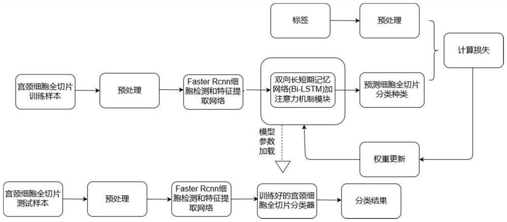
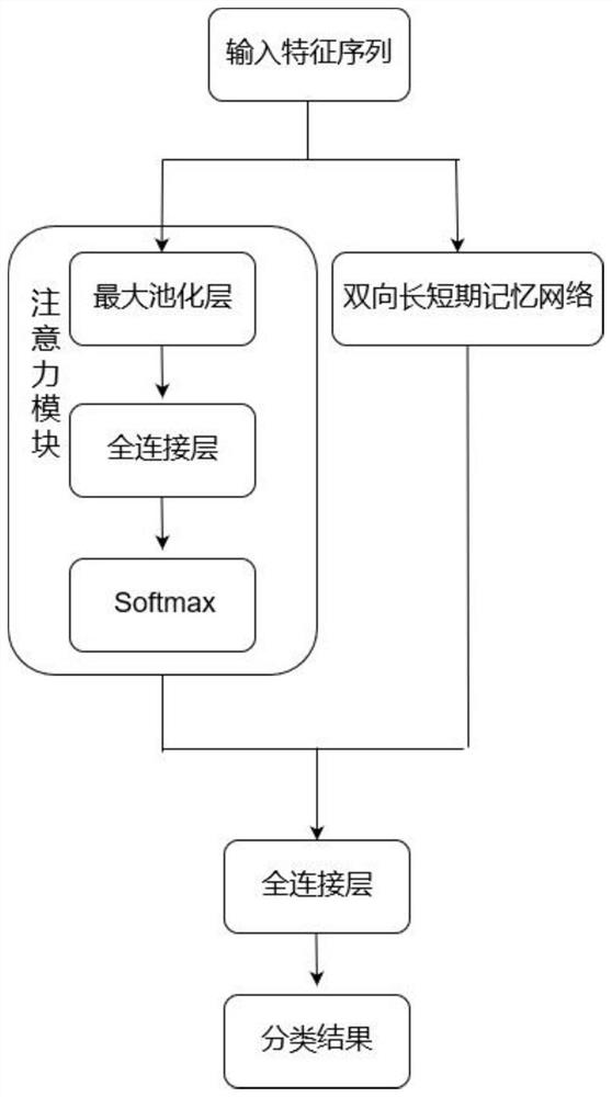
[0035] In this embodiment, a method for classifying cervical cytopathology full slices based on context modeling mainly uses the Faster-Rcnn network to detect cells in the full slice and extract cell nucleus image unit features, and uses a bidirectional long-short-term memory network ( Bi-LSTM) and the attention mechanism are used to model the image features of the whole slice, learn the features, and perform the classification prediction of the whole slice of cervical cells (wsi) through the cervical cell whole slice (wsi) classifier, such as figure 1 As shown, the specific steps are as follows:

[0036] Step 1. Obtain the whole slice sample of cervical cells with the dimension of H×W×C in class T and perform normalized preprocessing to obtain the preprocessed whole slice sequence and use it as a training sample, denoted as S={S 1 ,S 2 ,...,S t ,...,S T}, where S t Denotes the normalized cervical cell slice sample of the tth class, and Indicates the normalized cervica...


PUM


Abstract

The invention discloses a cervical cell full slice classification method based on context modeling. The method comprises the following steps: 1, obtaining T-type cervical cell full slice samples; 2, constructing a Faster Rcnn network-based cell detection and feature extraction module, carrying out cell detection and feature extraction on the cervical cell full slice sample, and extracting features from cell nucleus images with fixed sizes to obtain feature sequences of the cell nucleus images in different types of cell full slices; 3, building a context modeling module fusing the bidirectional long-short-term memory network and the attention mechanism; 4, constructing a cervical cell full slice classifier; and 5, carrying out classification prediction on the cervical cell full slice. More effective learning is carried out on the information obtained after feature extraction is carried out on the cell nucleus images in the input different cervical cell full slices, accurate classification of multiple different cervical cell full slices is completed, and the cell level marking cost of a current cervical cell classification method can be effectively reduced.

Description

technical field [0001] The invention relates to the technical field of digital image processing, in particular to image classification technology, in particular to a method for classifying cervical cytopathological full slices based on context modeling. Background technique [0002] Traditional pathological image analysis is completed by pathologists manually reading under a microscope. This link requires doctors with rich clinical experience to analyze a large number of histopathological slice images. However, the number of scans that doctors read every day is very large. , The reading time is long, and doctors may seriously affect the accuracy of pathological slide analysis due to overloaded workload and subjective emotions, which has brought great obstacles to the development of pathological slide analysis. If it is possible to improve the automatic process of cervical cell slice classification and use computer-aided film reading, it will bring new development ideas to pa...

Claims


Application Information

Patent Timeline
IPC IPC(8): G06K9/00G06K9/32G06K9/46G06K9/62G06N3/04G06N3/08
CPCG06N3/049G06N3/08G06N3/045G06F18/213G06F18/241
Inventor 刘波史骏唐昆铭束童祝新宇罗庭辉张元郑利平
Owner HEFEI UNIV OF TECH
Why Eureka
Try Eureka