Automatic segmentation of anatomical structure of large area annular ablation points

An anatomical structure, large-area technology, applied in medical automation diagnosis, image analysis, parts of surgical instruments, etc., can solve problems such as WACA limitation of ablation lines

Pending Publication Date: 2022-02-18
伯恩森斯韦伯斯特(以色列)有限责任公司
View PDF0 Cites 0 Cited by
  • Summary
  • Abstract
  • Description
  • Claims
  • Application Information

AI Technical Summary

Problems solved by technology

However, WACA is limited by potential gaps between ablation points, pot...

Method used


Image

  • Automatic segmentation of anatomical structure of large area annular ablation points
  • Automatic segmentation of anatomical structure of large area annular ablation points
  • Automatic segmentation of anatomical structure of large area annular ablation points

Examples


Embodiment Construction

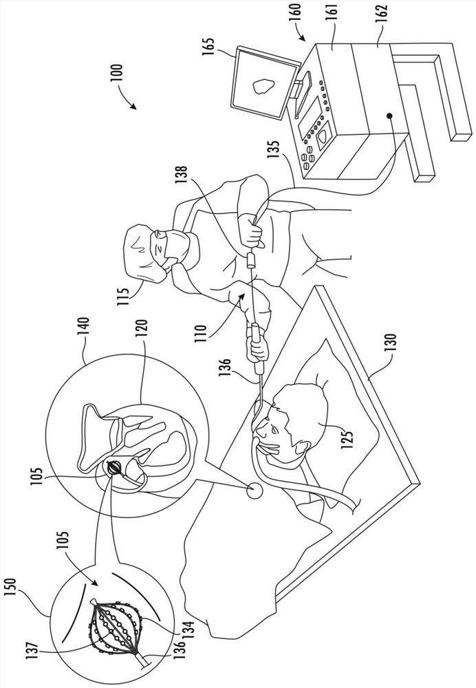
[0028] Disclosed herein is a system and method for automatic segmentation of the anatomy of a large area circular ablation (WACA) site. The systems and methods include artificial intelligence and machine learning. More specifically, the present disclosure relates to a system and method for automatic segmentation of anatomical structures of WACA points, the system and method including machine learning algorithms that perform efficient WACA ablation and efficient ablation during the cardiac mapping phase Automatic segmentation of the anatomy of points (eg, valid points). For example, the systems and methods include processor-executable code or software that resides in the process operation of the medical device device and resides in the processing hardware of the medical device device to use random forest regression, fully connected dense layers, and convolutional neural networks ( CNN) architecture to perform automatic segmentation of the anatomy of valid points.

[0029] Acc...


PUM


Abstract

The invention relates to automatic segmentation of anatomical structures of large area annular ablation points. A method and apparatus for implementing an evaluation engine implemented using a processor coupled to a memory is provided. The evaluation engine receives valid points corresponding to cardiac tissue of a patient. The evaluation engine determines an anatomical classification for each of the valid points based on a structural segmentation of the cardiac tissue, and provides the anatomical classification with each of the plurality of valid points to support treatment of the cardiac tissue.

Description

[0001] Cross References to Related Applications [0002] This application claims the benefit of US Provisional Patent Application 63 / 059,060, filed July 30, 2020, which is incorporated by reference as if fully set forth. technical field [0003] The present invention relates to methods and systems for artificial intelligence and machine learning. More specifically, the present invention relates to systems and methods utilizing machine learning algorithms that perform automatic segmentation of anatomy at large area circular ablation (WACA) points. Background technique [0004] Treatment of cardiac disorders often requires cardiac imaging (i.e., imaging of cardiac tissue, chambers, veins, arteries, and / or pathways, also known as cardiac scan or cardiac imaging) and subsequent ablation (i.e., removal or destruction of the imaged cardiac tissue, chambers, veins, arteries and / or pathways). In an example, a cardiac condition of atrial fibrillation may be imaged and treated usin...

Claims


Application Information

Patent Timeline
IPC IPC(8): A61B18/12
CPCA61B18/12A61B2018/00904A61B2018/00839A61B2018/00577A61B2018/00351A61B2018/00875A61B5/053A61B5/287A61B18/1492G16H50/20A61B5/7264A61B5/7267G06T7/11G06T7/155G06T2207/10028G06T2207/20081G06T2207/20084G06T2207/20092G06T2207/30048
Inventor S·戈德堡M·阿米特Y·A·阿莫斯
Owner 伯恩森斯韦伯斯特(以色列)有限责任公司
Why Eureka
Try Eureka