Automatic segmentation of tissues by dynamic change characterization

A technology of image segmentation and segmentation steps, applied in image analysis, instruments for radiological diagnosis, image data processing, etc., can solve the problem of difficult segmentation of tissues of interest or other substances

Inactive Publication Date: 2007-02-14
KONINKLIJKE PHILIPS ELECTRONICS NV
View PDF0 Cites 3 Cited by
  • Summary
  • Abstract
  • Description
  • Claims
  • Application Information

AI Technical Summary

Problems solved by technology

[0006] While the techniques described above are effective, tissue of interest or other matter remains difficult to segment even with a combination of a contrast-enhanced image and a corresponding image without contrast agent

Method used


Image

  • Automatic segmentation of tissues by dynamic change characterization
  • Automatic segmentation of tissues by dynamic change characterization
  • Automatic segmentation of tissues by dynamic change characterization

Examples


Embodiment Construction

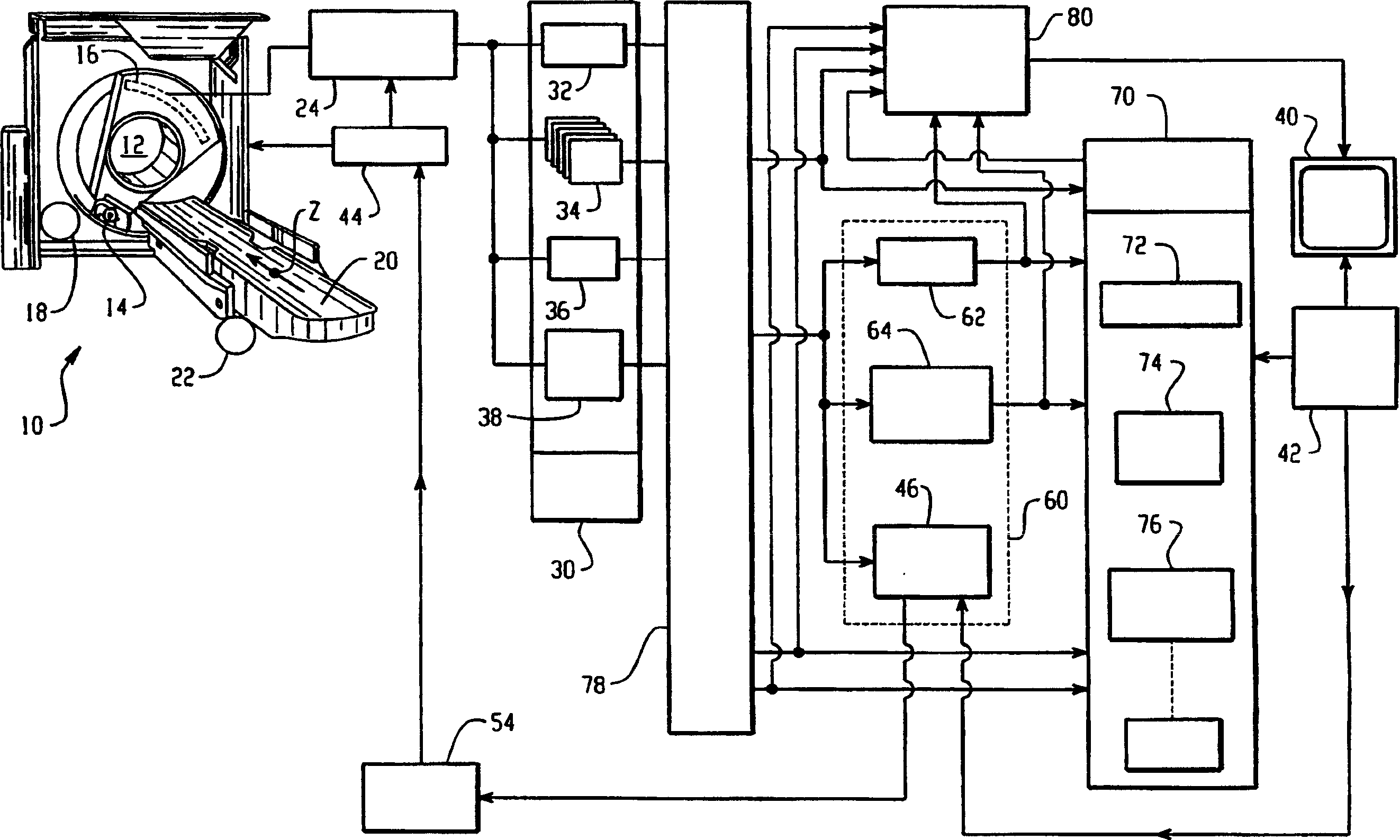
[0015] refer to figure 1 , a diagnostic imaging apparatus, such as a CT scanner 10, non-invasively examines a subject located in an imaging region 12 and generates diagnostic image data representing a region of interest of the subject placed in the examination region. In the described CT scanner embodiment, an x-ray tube 14 or other radiation source generates a beam, preferably a multilayer or cone beam, which is directed towards a radiation detector 16 through an examination region 12 . A motor 18 and associated drives (not shown) rotate the x-ray beam around the examination region, typically by rotating the x-ray source and detectors. The subject is supported on a patient support 20 such as a subject couch. A motor 22 and associated engagement and coupling means (not shown) propels the subject longitudinally through the examination region such that the subject's region of interest passes through the examination region. For a helical scan, while the motor 18 rotates the x-r...


PUM


Abstract

A reconstruction processor(24) reconstructs diagnostic data from a diagnostic imaging device, such as a CT scanner(10), starting before a contrast agent reaches a region of interest(50), as the concentration of contrast agent in the region of interest builds(52), and at a contrast agent peak(56). The plurality of images generated while the contrast agent concentration is building are aligned(78). A change map is generated indicative of a rate-of-change(62) gradient or a time-to-peak(64) for corresponding pixels or voxels of the images generated during the time the contrast agent is building to the peak. A segmentation processor(70) uses the change map in segmenting the diagnostic images generated without contrast agent or at the contrast agent concentration peak.

Description

technical field [0001] The present invention relates to diagnostic imaging techniques. The present invention finds particular application in relation to the segmentation of contrast-enhanced diagnostic images into different tissue regions or organs, and will be described in detail herein. Background technique [0002] In many diagnostic imaging situations, a diagnostician attempts to determine the boundaries between different tissues of interest, such as blood and the walls of blood vessels. Typically, diagnosticians rely on differences in intensity or grayscale to distinguish different tissues. Various computer programs have been developed to automatically segment or distinguish different tissues in diagnostic images, rather than relying on the human eye to make such distinctions. [0003] Often, it is quite difficult to distinguish tissues of interest, plaques, and other structures by intensity or grayscale. Especially when one tissue is blood, one technique facilitates...

Claims


Application Information

Patent Timeline
Patent Type & Authority Applications(China)
IPC IPC(8): G06T7/00G06T5/00
CPCG06T2207/10081A61B6/481G06T2207/20104G06T2207/10121G06T7/0012G06T7/2006G06T2207/30101G06T2207/10076A61B6/504G06T7/215
Inventor O·海A·马科维奇I·莱夫
Owner KONINKLIJKE PHILIPS ELECTRONICS NV
Why Eureka
Try Eureka