Device for three dimensional endoscopic surgery

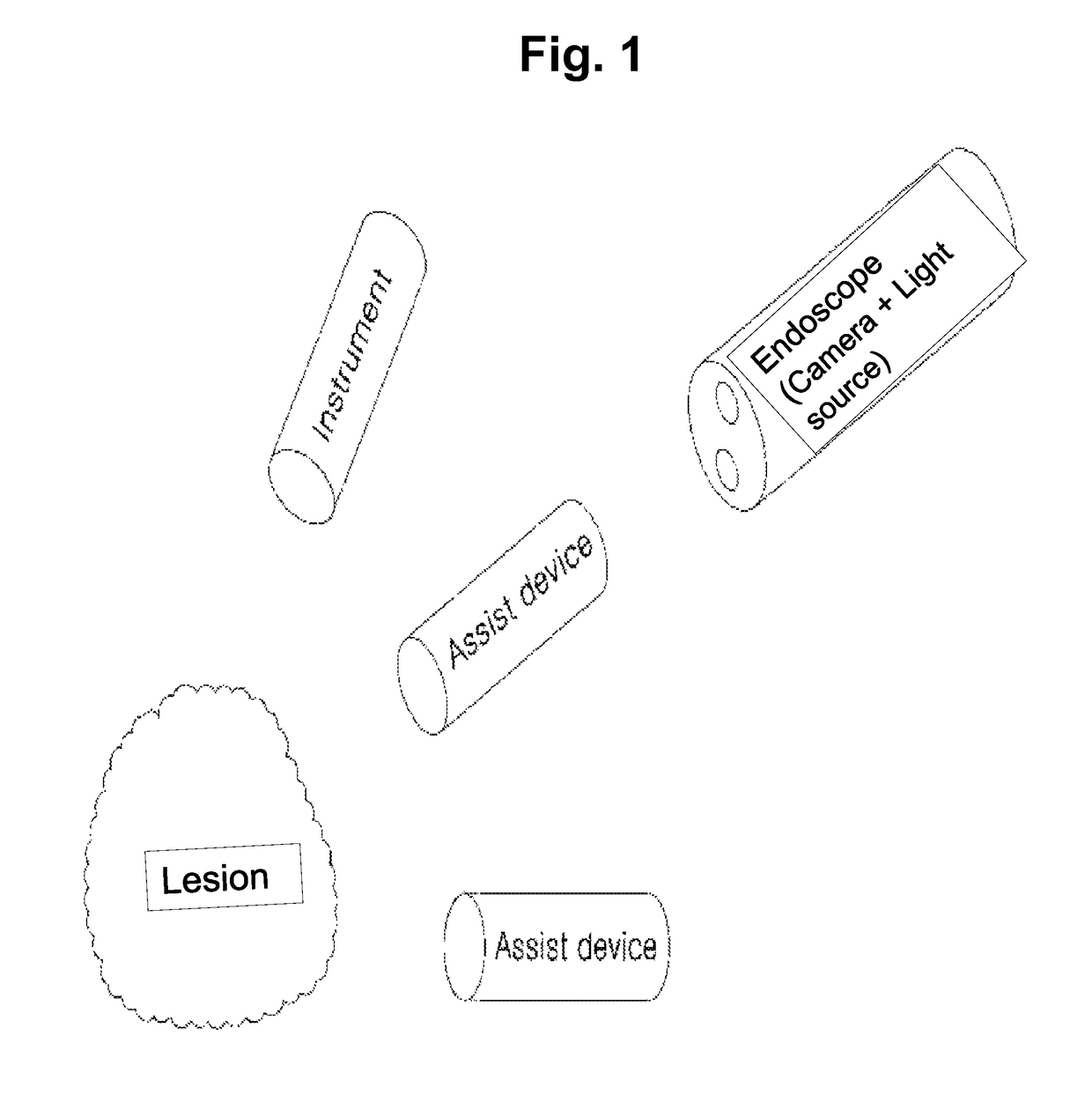
a three-dimensional endoscopic and surgery technology, applied in the field of three-dimensional endoscopic surgery, can solve the problems of increasing the area to be incised at the time of surgery, the incision window through which the instruments necessary for treatment and surgery can be inserted needs to be further formed, and the need for four incision windows is needed. to achieve the effect of clear three-dimensional images

Active Publication Date: 2018-07-24
CATHOLIC KWANDONG UNIV IND FOUND
View PDF15 Cites 1 Cited by
  • Summary
  • Abstract
  • Description
  • Claims
  • Application Information

AI Technical Summary

Benefits of technology

[0009]Therefore, the present invention has been made in view of the above-mentioned problems, and an aspect of the present invention is to provide a device for three-dimensional endoscopic surgery in which therapeutic instruments including an endoscope can be inserted into the human body through one main tube and can be spread outward to be utilized in the human body, thereby enabling surgery through a natural opening such as a nasal cavity or throat, minimizing an incision site at the time of surgery, and performing various medical works using one main tube.
[0010]Another aspect of the present invention is to provide a device for three-dimensional endoscopic surgery in which various functional instruments are inserted into the human body using one main tube, thereby requiring no additional incision window.
[0012]Still another aspect of the present invention is to provide a device for three-dimensional endoscopic surgery in which a large camera is installed inside a main tube, and washing modules for washing cameras and a light source are provided inside the main tube, thereby providing clearer three-dimensional images during performing of medical works.
[0026]Here, locking devices may be connected to the first to fifth shafts for preventing rotation of the first to fifth shafts.
[0029]According to the present invention, therapeutic instruments including an endoscope can be inserted into the human body through one main tube and can be spread outward to be utilized in the human body, thereby minimizing an incision site at the time of surgery, utilizing natural openings without separate incision windows, and performing various medical works using one main tube.
[0031]Further, according to the present invention, large-diameter cameras can be installed inside a main tube, and washing modules for washing cameras and a light source can be provided inside the main tube, thereby providing clearer three-dimensional images during performing of medical works

Problems solved by technology

However the conventional laparotomy has a disadvantage in that a total of four incision windows are needed since an endoscope for photographing a lesion site, an instrument for performing surgery, and assisting devices need to be respectively inserted, as shown in FIG. 1.
In other words, although the recent medical demand is that the cross-sectional area of the endoscope is made as small as possible so that diagnosis or treatment is performed using a natural opening (nasal cavity or throat) or a small incision window in a non-invasive or less invasive manner, the foregoing method has a disadvantage in that the area to be incised is increased at the time of surgery.
The foregoing configuration has an advantage in that two photographing parts and an illuminating parts for photographing three-dimensional images are folded inside the pipe-shaped body and the photographing parts are spread in the human body, so that the diameter of the body is decreased, thereby minimizing the incision at the surgical site, but has a disadvantage in that an incision window through which instruments necessary for treatment and surgery can be inserted needs to be further formed.
The foregoing configuration also has a disadvantage in that, since a separate device for washing the photographing parts is not prepared, a photographed image cannot be differentiated in cases where foreign substances are attached on lenses of the photographing parts inserted into the body.

Method used


Image

  • Device for three dimensional endoscopic surgery
  • Device for three dimensional endoscopic surgery
  • Device for three dimensional endoscopic surgery

Examples


Embodiment Construction

[0046]Hereinafter, preferable embodiments of a device for three-dimensional endoscopic surgery according to the present invention will be described in detail with reference to the accompanying drawings.

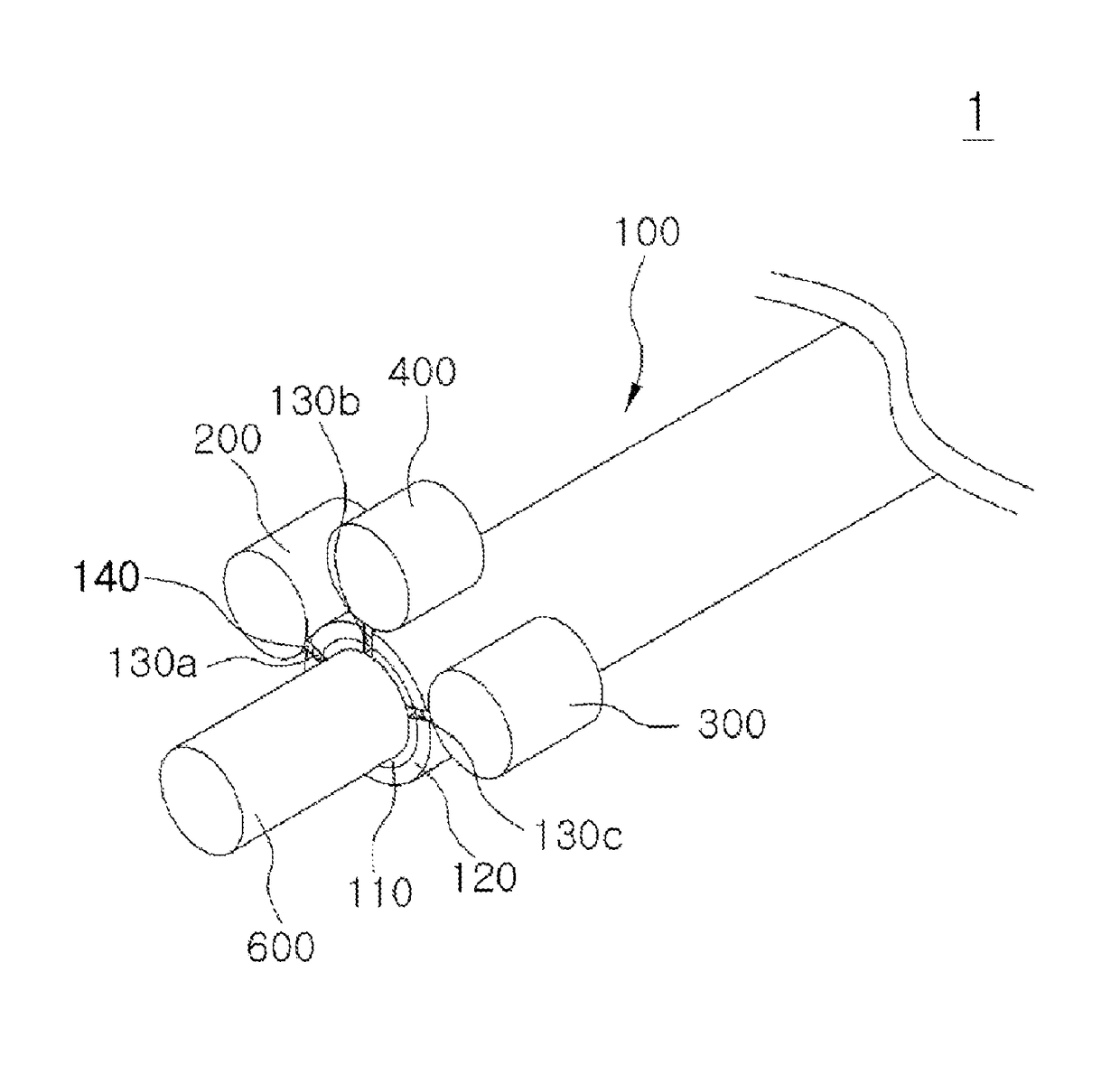
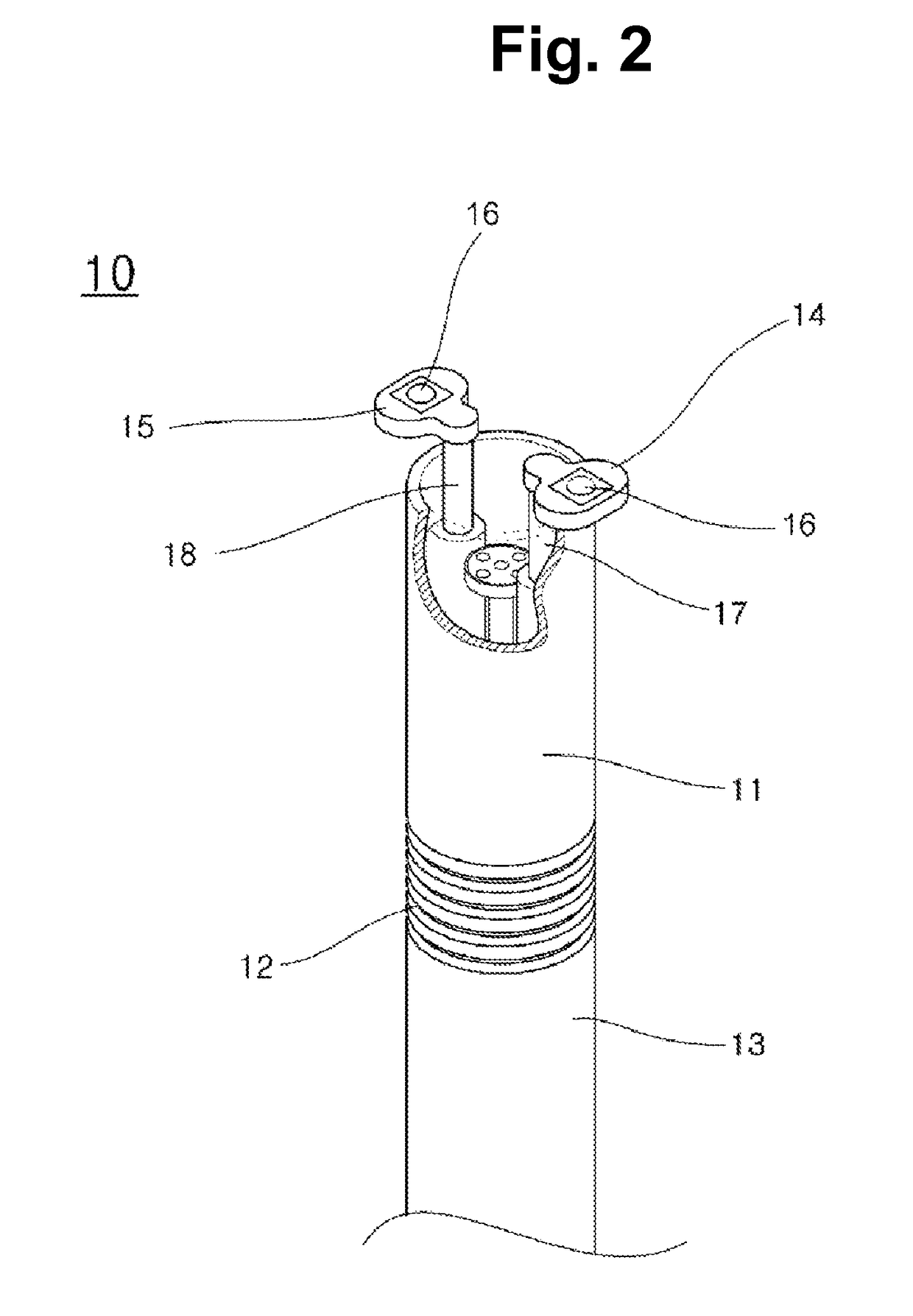
[0047]FIG. 3 is a perspective view showing a device for three-dimensional endoscopic surgery according to an embodiment of the present invention; FIG. 4 is a perspective view showing a device for three-dimensional endoscopic surgery according to another embodiment of the present invention; FIG. 5 is a partial cross-sectional view showing a main tube and an inner structure thereof; FIGS. 6(a) and (b) are partial cross-sectional views schematically showing a structure of a main tube in a device for three-dimensional endoscopic surgery according to the present invention; FIGS. 7 and 8 are perspective views illustrating an operative relationship of a device for three-dimensional endoscopic surgery according to the present invention; FIG. 9 is a cross-sectional view schematically showing i...


PUM


Abstract

The present invention relates to a device for three dimensional endoscopic surgery, and more specifically, to a device for three dimensional endoscopic surgery, wherein therapeutic tools including an endoscope can be inserted into the human body through one main tube, and the therapeutic tools can be spread outward to be utilized in the human body, thereby minimizing an incision site in case of surgical operation while allowing various medical operations to be performed using one main tube. The present invention is a device for endoscopic surgery comprising: a main tube which is inserted into the body of a patient; first and second cameras which are inserted in a row into the main tube; a light source portion which is inserted between the first and second cameras; and a therapeutic tool which is located below the second camera to be inserted into the main tube.

Description

TECHNICAL FIELD[0001]The present invention relates to a device for three-dimensional endoscopic surgery, and more specifically, to a device for three-dimensional endoscopic surgery in which surgical instruments including an endoscope can be inserted into the human body through one main tube and can be spread outward to be utilized in the human body, thereby minimizing an incision site at the time of surgery and performing various medical works using one main tube.BACKGROUND ART[0002]In general, an endoscope refers to a medical instrument which is used to directly look inside internal organs or body cavities, and is an instrument which has been made to be inserted into an organ of which a lesion cannot be directly observed without surgery or autopsy and enables observation of the lesion.[0003]In order to solve problems of the conventional laparotomy in which an abdominal site is incised with a knife, the endoscopy operation has been recently developed and conducted such that small ho...

Claims


Application Information

Patent Timeline
Patent Type & Authority Patents(United States)
IPC IPC(8): A61B1/04A61B1/313A61B1/00A61B90/00A61B1/12A61B17/00A61B90/30A61B1/05A61B1/06A61B34/30
CPCA61B1/3132A61B1/00128A61B1/00181A61B1/00193A61B1/051A61B1/0676A61B1/126A61B17/00234A61B90/30A61B90/361A61B90/37A61B1/00087A61B2090/371A61B34/30A61B2017/0034A61B2017/00296A61B2034/302A61B2090/309A61B1/0014A61B1/00188A61B1/00194A61B17/00A61M25/01A61B1/05A61B1/06
Inventor YOON, CHEESOON
Owner CATHOLIC KWANDONG UNIV IND FOUND
Why Eureka
Try Eureka