Systems and methods for detection of structures and/or patterns in images

a system and image technology, applied in the field of image analysis, can solve the problems of limiting the detection technique to automatic nucleus rather than membrane detection, subject to intra- and inter-individual variability, and reducing so as to reduce the computational burden, increase the stability of the cnn, and reduce the number of training images

Active Publication Date: 2018-10-23
VENTANA MEDICAL SYST INC
View PDF5 Cites 21 Cited by
  • Summary
  • Abstract
  • Description
  • Claims
  • Application Information

AI Technical Summary

Benefits of technology

[0018]Embodiments of the invention are particularly advantageous as a convolutional neural network is employed for generating a probability map representing a probability for the presence of the biological features that has a structure which facilitates the training of the convolutional neural network (CNN), provides enhanced stability and reduces the computational burden and latency times experienced by the user. This is accomplished by connection mapping of the inputs of the CNN to feature maps of its first convolutional layer such that subsets of the channels that are representative of co-located biological features are mapped to a common feature map. By using the a priori biological knowledge as regards the co-location of stains a structure is thus enforced onto the CNN that has these advantageous effects. This is done by a step of configuring the CNN correspondingly.
[0019]In accordance with an embodiment of the invention the number of feature maps is below the number of channels of the multi-channel image. This is particularly advantageous for reducing the computational burden and increased stability of the CNN as well as to reduce the number of training images that are required for training the CNN.
[0020]In accordance with a further embodiment of the invention the image sensor that is used to acquire the multi-channel image has a number of color channels that is below the number of channels of the multi-channel image. The co-location data that describes the co-location data that describes the co-location of stains may be utilized for performing the unmixing, such as by using a group sparsity model as it is as such known from the prior art. This way the co-location data can be used both for performing the unmixing and for configuring the CNN.
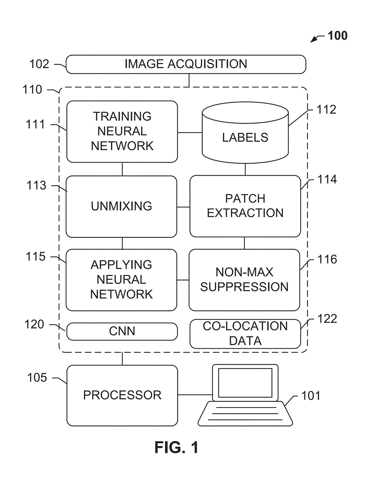
[0021]The subject disclosure solves the above-identified problems by presenting systems and computer-implemented methods for automatic or semi-automatic detection of structures of interest within images, for example, cellular structures (e.g., cells. nuclei, cell edges, cell membrane), background (e.g., background patterns such as white or white-like space), background image components, and / or artifacts. In exemplary embodiments of the present invention, the present invention distinguishes cellular structures in an image from non-cellular structures or image components. The structures or components may be identified using a convolutional neural network that has been trained for this task. More particularly, the convolutional neural network may be trained to recognize specific cellular structures and features using training images and labels. The neural network outputs a probability that the detected structure does in fact represent a cell, membrane, background, etc. These probabilities may undergo a local maxima finding method such as non-maximum suppression in order to identify a particular pixel that will be used as the “location” of the object. A particular part of the cell, e.g., the approximate center of a nucleus, is illustratively used as the “location” of the object within the area under observation, i.e. an image patch.
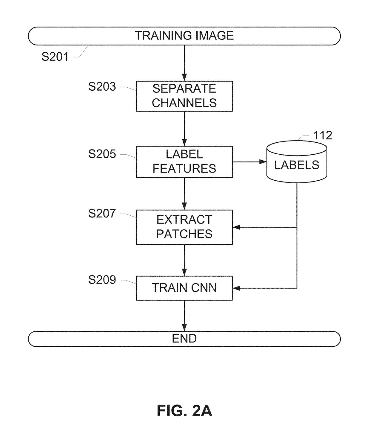
[0022]Operations described herein include retrieving individual color channels from a multi-channel image and providing said multiple individual channels as input for a detector, for example, a cell detector. The cell detector may comprise a learning means that is trained using ground truths for cellular structures, such as cells, portions of cells, or other cell or image features identified by a trained operator, such as a pathologist. The trained cell detector may be used to identify cellular structures, such as immune cells, in the channels of the image that correspond to multiple types of cell markers or other target structures such as a nucleus. The learning means may include generating a convolutional neural network (CNN) by analyzing a plurality of training images with ground truths labeled thereon. Subsequent to the training, a test image or image under analysis may be divided into a plurality of patches, each patch containing one or multiple channels that are classified according to a CNN, and a probability map may be generated representing a presence of the immune cell or other target structure within the image. Further, a non-maximum suppression operation may be performed to obtain the coordinates of the target structure from the probability map.
[0023]In exemplary embodiments described herein, multiple types of cells, for example, immune cells may be detected from a multi-channel image, such as an original RGB image acquired from a brightfield imaging system, an unmixed fluorescent image, or an image in any other color space such as LAB. In alternate exemplary embodiments described herein, the detection can be applied to selected regions of the image instead of the whole image, and for example, enabled by detecting the foreground of the image, and only apply detection within the foreground region. To accelerate this cell detection process, a precomputed foreground mask can be used to enable processing of only regions of the image that are likely to contain immune cells in their foreground.

Problems solved by technology

This is an extremely tedious and time consuming process and the results may also subject to intra- and inter-individual variability.
However, these techniques are limited to automatic nucleus detection rather than membrane detection.
Although some machine learning based systems use scale invariant feature transform (SIFT) for maintaining sufficient contrast of cell boundaries, this method was developed for unstained cell images and it is non-trivial to extend it to detect immune cells in IHC stained images.

Method used


Image

  • Systems and methods for detection of structures and/or patterns in images
  • Systems and methods for detection of structures and/or patterns in images
  • Systems and methods for detection of structures and/or patterns in images

Examples


Embodiment Construction

[0036]The subject disclosure solves the above-identified problems by presenting systems and computer-implemented methods for automatic detection of image structures, for example, cellular structures, including retrieving individual color channels from a multi-channel image and providing one or multiple individual channels or portions of image data from the one or more multiple individual channels as input for a cell detector that is trained using a convolutional neural network to identify the immune cells in one or multiple channels of the image that corresponds to an immune cell marker or other target structure such as a nucleus. The multi-channel image may be an RGB image obtained from a brightfield scanner, an image from another color space such as Lab, a multi-channel image from a multi-channel brightfield or darkfield scanner, a fluorescent image from a multi-spectral imaging system, a darkfield image, or any other multi-channel image. In some embodiments the image may be an im...


PUM


Abstract

The subject disclosure presents systems and computer-implemented methods for automatic immune cell detection that is of assistance in clinical immune profile studies. The automatic immune cell detection method involves retrieving a plurality of image channels from a multi-channel image such as an RGB image or biologically meaningful unmixed image. A cell detector is trained to identify the immune cells by a convolutional neural network in one or multiple image channels. Further, the automatic immune cell detection algorithm involves utilizing a non-maximum suppression algorithm to obtain the immune cell coordinates from a probability map of immune cell presence possibility generated from the convolutional neural network classifier.

Description

RELATED APPLICATIONS[0001]This patent application is a continuation of International Patent Application No. PCT / EP2015 / 061226 filed May 21, 2015, which claims priority to and the benefit of U.S. Provisional Application No. 62 / 098,087, filed Dec. 30, 2014, and entitled “SYSTEMS AND METHODS FOR DEEP LEARNING FOR CELL DETECTION”, and to U.S. Provisional Application No. 62 / 002,633, filed May 23, 2014, and entitled “DEEP LEARNING BASED AUTOMATIC CELL COUNTING SYSTEM AND METHOD”, the contents of which are hereby incorporated by reference herein in their entirety into this disclosure.BACKGROUND OF THE SUBJECT DISCLOSURE[0002]Field of the Subject Disclosure[0003]The present subject disclosure relates to image analysis. More particularly, the present subject disclosure relates to automatically identifying structures (e.g., cellular structures) or patterns (e.g., background or white space) in an image.[0004]Background of the Subject Disclosure[0005]In the analysis of biological specimens such...

Claims


Application Information

Patent Timeline
Patent Type & Authority Patents(United States)
IPC IPC(8): G06K9/00G06T7/00
CPCG06T7/0012G06K9/00127G06T2207/30024G06T2207/20081G06T2207/20084G06T2207/10024G06N3/084G06V20/69G06V20/698G06N3/045
Inventor CHEFD'HOTEL, CHRISTOPHECHEN, TING
Owner VENTANA MEDICAL SYST INC
Why Eureka
Try Eureka