Near-infrared spectrum imaging system and method for diagnosis of depth and area of burn skin necrosis

a skin necrosis and near-infrared technology, applied in the field of tissue injury and spectral imaging technology, can solve the problems of increasing the depth of the burn, affecting the safety and health of the body, and affecting the health of the patient, and achieves high efficiency, wide field of view angle, and high resolution.

Active Publication Date: 2019-05-07
BEIJING HEFENGLIANKANG INVESTMENT MANAGEMENT LTD
View PDF1 Cites 16 Cited by
  • Summary
  • Abstract
  • Description
  • Claims
  • Application Information

AI Technical Summary

Benefits of technology

[0014]The present invention is made with respect to the above defects existing in the prior art and aims to provide a near infrared spectrum imaging system and method for the diagnosis of burn skin necrosis depth and area, which uses super spectrum imaging instrument with high resolution, wide field of view angle, and high efficiency to obtain the key information of degeneration and necrosis tissue of burn skin by non-contact and noninvasive method and is visible. It obtained the accurate information of burn skin necrosis area and depth by information processing; and providing key information about the necrotic tissue of skin burn to clinicians for clinical treatment and clinical surgery.
[0033](3.1) spectrum correction: the amplitude of spectrum curve corresponding to each image pixel of the spectral image in the original spectral image of the target region is divided by the amplitude of spectral curve corresponding to the image pixel of the spectral image of the white board in the same condition so as to remove the influence of the background light and the heterogeneity of the light source, and the spectral reflectance curve of the target region is obtained;
[0036]The imaging system and method provided by the present invention as above have the characteristics of high resolution, wide field of view angle and high efficiency, and is capable of indicating the changes of the spatial structure of the skin caused by the degeneration of tissue protein before and after the skin burn. Therefore, the micron level information about the boundary between normal skin tissue and necrotic tissue and the depth of burn skin necrosis are provided in a non-contact and noninvasive manner and the visualization is achieved. Finally, the key information of necrosis depth and area of the skin burn and necrosis tissue in micron level was provided to clinicians, which can be beneficial to guide clinical therapy and clinical surgery and to avoid cross infection and patient pain caused by medical detection to the maximum extent. It is beneficial for clinical diagnosis, treatment and prognosis.

Problems solved by technology

After burn, the mortality rate, appearance damage rate and mutilation rate are high, the treatment period is long, which directly threaten the life safety and the health of the body, which brings heavy burden to the society and the family.
At present, the accurate judgment of the degree of skin tissue damage caused by burns is still a major problem in burn surgery, but the most common method used in clinical diagnosis of burn depth still mainly relies on clinical experience, including the observation of the appearance of the burn wound, the condition of capillary refill, and the feeling of the wound on the touch and the pain of acupuncture, etc.
The latter can increase the depth of the burn, that is, wound may become deeper.
In addition, deep burn skin tissue lesions can be deep into the muscle and even bone, which is far beyond the anatomical depth of the skin tissue, and the treatment and prognosis of this kind of injury are more complicated, therefore, the empirical diagnosis of burn depth only based on the naked eye identification has a limited guiding effect for the clinical treatment.
There are several reasons why this cannot be applied to clinical practice: 1. the operation of a biopsy is traumatic to the body, which is not acceptable to some patients; 2. in a certain period of time, the pathological changes of the burned tissue are dynamic and continuous, therefore, it may not accurately predict the results by performing a single biopsy so as to preliminarily assess the damage degree in the early stage of the burn; 3. an experienced pathology expert is required, and this expert has to commit to this work for a long time.
In addition, the fluorescence is weak when the deep burn blood vessel is damaged or is blocked, which increase detection error.
However, this method has higher requirements for the detection condition, and needs constant ambient temperature and equilibrium time, both evaporation and cooling of wounds greatly affect the detection results.
Due to high equipment cost, large individual difference, high false positive and many other factors, Deep II and III degree wounds cannot be well distinguished, limiting its wide application in clinical.
The main factor that limits its clinical application is that contact of the wound during the operation is required.
In addition, practice has proved that the diagnostic accuracy is low, and is not superior to the subjective empirical diagnosis.
However, the core principles of the above research are to judge the depth of burn indirectly based on the changes of oxygen carrying status of red blood cell in blood, and tissue water content changes of the skin before and after skin burn (see FIG. 9), and precise information about the depth and area of burn skin tissue necrosis cannot be provided directly.
At present, its imaging band is mainly in the visible and near infrared spectrum, but the spectral resolution is generally low, and the scanning precision is about 50 nm, which cannot meet the needs of medical fine research yet.
The existing spectral imaging techniques can not accurately distinguish the skin necrosis tissue and acquire the spectral images of the skin tissue tomography, there is also a key point that the spectral resolution and imaging spectral range thereof cannot distinguish skin necrotic tissue and acquire the tomography images thereof, that is, a high resolution narrow band and wide band tunable filter is the core problem.
Therefore, so far, there is no such diagnostic equipment and related methods which can directly and accurately distinguish the depth of the necrotic tissue, accurately identify the boundaries of the necrotic tissue and normal tissue.

Method used


Image

  • Near-infrared spectrum imaging system and method for diagnosis of depth and area of burn skin necrosis
  • Near-infrared spectrum imaging system and method for diagnosis of depth and area of burn skin necrosis
  • Near-infrared spectrum imaging system and method for diagnosis of depth and area of burn skin necrosis

Examples


Embodiment Construction

[0054]In order to better illustrate and explain the technique of the present invention and its effects, the present invention is described in further detail below with reference to the drawings and research experiment process of the present invention.

[0055]I. The Clinical Diagnosis System for the Depth and the Area of Burn Skin Necrosis

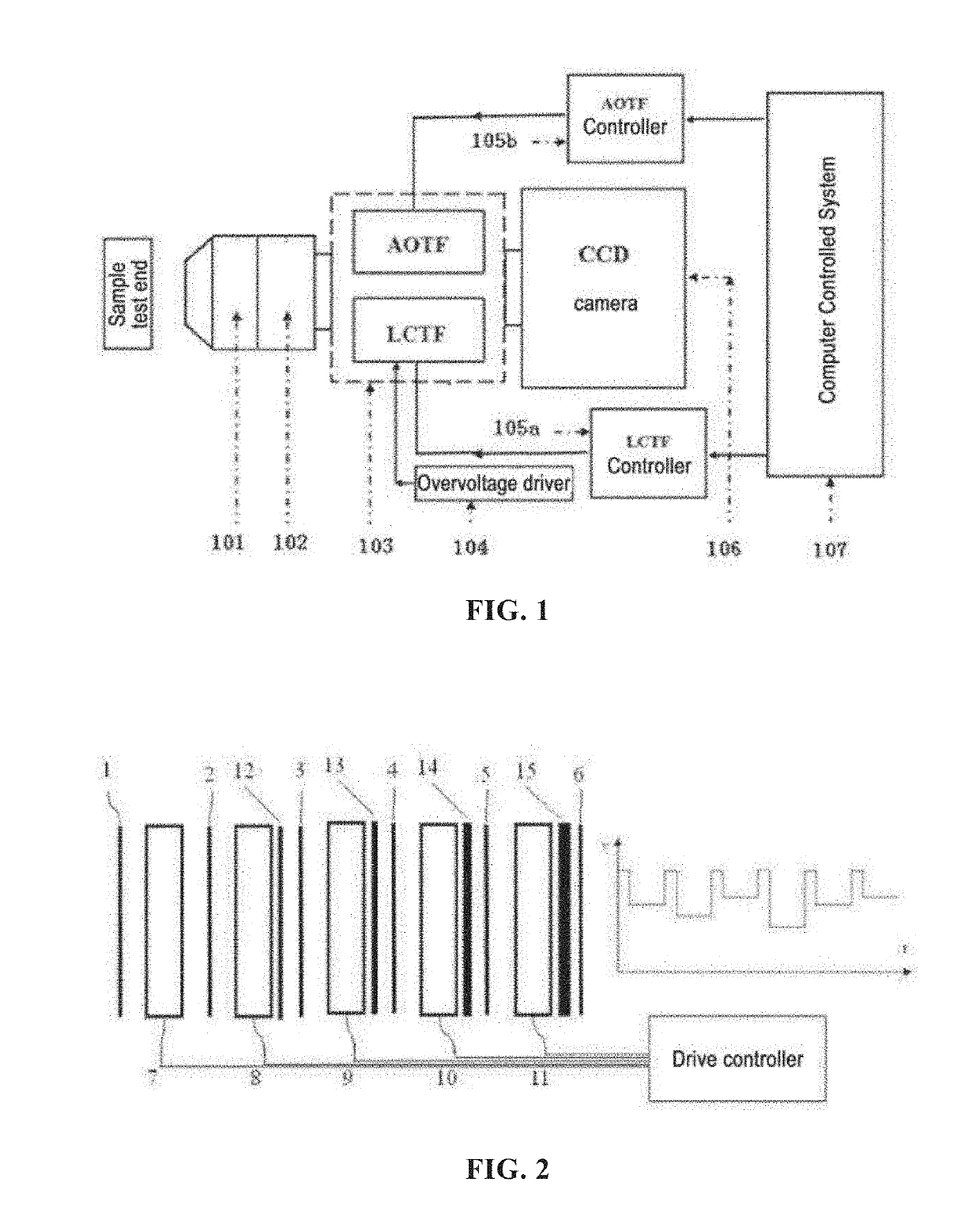
[0056]The diagnosis system mainly includes the spectral imaging instrument and the computer control system. The structure is shown in FIG. 1, wherein:

[0057]1. The spectral imager: consisting of an illumination light source101, an optical lens 102, a filter 103 (using a liquid crystal tunable filter LCTF and an acousto-optic tunable filter AOTF), a CCD camera 106, a LCTF controller 105a and AOTF controller 105b and an overvoltage driver 104. They are assembled in the form of a conventional spectral imager. The performance parameters of each component can be selected as follows:

[0058]Liquid crystal tunable filter (LCTF): working band: 900 nm˜2500 nm; sp...


PUM


Abstract

A near-infrared spectrum imaging system for diagnosis of the depth and the area of burn skin necrosis comprises a spectrum imager and a computer controlled system. The spectrum imaging system comprises a light source (101), an optical lens (102), a filter (103), a driving controller (105a, 105b, 104), and a CCD camera (106). The filter (103) uses a wide-spectrum liquid crystal tunable filter (LCTF) or an acousto-optic tunable filter (AOTF) that obtain 1100-2500 nm waveband spectral signals of burn skin necrosis tissue of a target region. A compute controlled system is internally provided with a universal module, a data module, a spectrum correction module, a spectrum matching module, and a burn wound three-dimensional synthesizing module. The spectrum imager obtains spectral image data of burn skin necrosis tissue of a target region and inputs the data into the computer controlled system, and the computer controlled system performs image analysis and processing of the data; the depth and the area of burn of the target region can be obtained by means of spectral matching and recognition on a spectral reflectance curve corresponding to each image pixel in an spectral image and a standard spectral reflectance curve in a burn skin necrosis spectral database in a data module, and the data is synthesized into a three-dimensional image for display.

Description

FIELD OF THE INVENTION[0001]The present invention relates to the field of tissue injury and spectral imaging technology.BACKGROUND OF THE INVENTION[0002]Burns are common diseases in peacetime and wartime, especially in wartime. After burn, the mortality rate, appearance damage rate and mutilation rate are high, the treatment period is long, which directly threaten the life safety and the health of the body, which brings heavy burden to the society and the family.[0003]At present, the accurate judgment of the degree of skin tissue damage caused by burns is still a major problem in burn surgery, but the most common method used in clinical diagnosis of burn depth still mainly relies on clinical experience, including the observation of the appearance of the burn wound, the condition of capillary refill, and the feeling of the wound on the touch and the pain of acupuncture, etc. Data shows that accuracy of the empirical diagnosis of wound surface by the world's best burn doctors is only ...

Claims


Application Information

Patent Timeline
Patent Type & Authority Patents(United States)
IPC IPC(8): A61B6/00A61B5/00
CPCA61B5/445A61B5/0075A61B5/7246A61B5/0077A61B2562/0242
Inventor WU, JUNLIU, CANGLIYANG, JUNJIELUO, YONGQUANHUANG, LIXIANLUO, GAOXINGCHEN, ZHIQIANGZHAO, JIANHENGZHANG, DAYONGSHEN, ZHIXUELI, JIANFENGHE, WEIFENG
Owner BEIJING HEFENGLIANKANG INVESTMENT MANAGEMENT LTD
Why Eureka
Try Eureka