
System and methods for treating MVO

a technology of system and method, applied in the field of system and method for treating mvo, can solve the problems of inability to diagnose microvascular obstruction in the cath lab, inability to treat mvo, shorten life,

Active Publication Date: 2019-06-11
CORFLOW THERAPEUTICS AG
View PDF9 Cites 11 Cited by
  • Summary
  • Abstract
  • Description
  • Claims
  • Application Information

AI Technical Summary

Benefits of technology

[0030]This application describes novel devices and methods for both diagnosing and treating MVO emergent in the Cath Lab. It allows monitoring treatment efficacy in real time during the therapeutic interventional procedure. The invention uses a compact computerized console and fluid pump with unique and specialized fluid infusion algorithms together with a unique catheter system having both diagnostic and therapeutic capabilities. MVO diagnosis can be made and treatment assessed in real time via flow controlled multi-agent (both therapeutic and diagnostic) infusion. The integrated system provides precise cardiac or other organ (brain, lung, kidney, limb, visceral organ and others) microcirculatory characterization in real-time to diagnose MVO and precisely guide therapy in real time.

Problems solved by technology

Large vessel occlusion is often associated with small vessel occlusion (termed Microvascular occlusion or MVO) by clot and embolic debris, also a serious problem since the heart muscle is deprived of blood, oxygen, and critical nutrients necessary to sustain cell life.
However, microvascular obstruction cannot be diagnosed in the Cath lab, and more importantly MVO cannot be treated even if / when it could be accurately diagnosed.
These complications can markedly shorten life and impose severe limitations on quality of life.
However, substantial challenges remain for treating STEMI patients and reducing long term complications.
These problems include heart failure (poor cardiac function and cardiac enlargement), cardiac / ventricular rupture, persistent ischemic chest pain / angina, left ventricular aneurysm and clot, and malignant arrhythmias.
Heart failure is worsened as the heart remodels in shape and size, and loses function.
Many years investigating STEMI therapy show that opening the epicardial / large coronary artery is insufficient to salvage heart muscle and optimize long term patient outcome.
Thus, despite a widely patent epicardial coronary artery, residual MVO obstructs blood flow into the heart causing cell ischemia death from severe heart muscle damage.
Too often, obstructed microvessels (MVO) do not resolve even after stent placement, and have serious long-term negative prognostic implications.
MRI, however, cannot be performed emergently or during a cardiac catheterization procedure since it requires patients to be in a separate imaging area and within a separate scanner. FIG. 13A shows regions of profound MVO at the myocardial infarct core, having very dark muscle with no contrast flowing into the cardiac segment. FIG. 13B is a repeat scan 6 months later in the same patient, and shows dense heart scarring (white line of tissue replacing the black region) that resulted from the MVO.
MVO cannot be diagnosed in a catheterization laboratory.
Moreover, no effective therapies are available.
Many possible prior therapies all proved essentially ineffective, and in some cases, dangerous.
Problems encountered with prior MVO therapy include rapid fluid bolus injection with drugs.
Delivering high drug doses to occluded microchannels in this adverse ratio yields unacceptably high toxic systemic drug level because all injected drug eventually enters the systemic circulation.
High systemic drug levels place patients at risk of dangerous systemic bleeding and other systemic complications including vessel dissection due to high flow infusion rates.
Technology and methods to successfully and efficiently deliver therapeutic agents to MVO-obstructed microvessels of multiple organs (Heart, brain, bowel, extremities, liver, kidneys for example) are not available.

Method used


Image

  • System and methods for treating MVO
  • System and methods for treating MVO
  • System and methods for treating MVO

Examples


Embodiment Construction

[0096]Specific embodiments of the invention will now be described with reference to the accompanying drawings. This invention may, however, be embodied in many different forms and should not be construed as limited to the embodiments set forth herein; rather, these embodiments are provided so that this disclosure will be thorough and complete, and will fully convey the scope of the invention to those skilled in the art. The terminology used in the detailed description of the embodiments illustrated in the accompanying drawings is not intended to be limiting of the invention. In the drawings, like numbers refer to like elements.

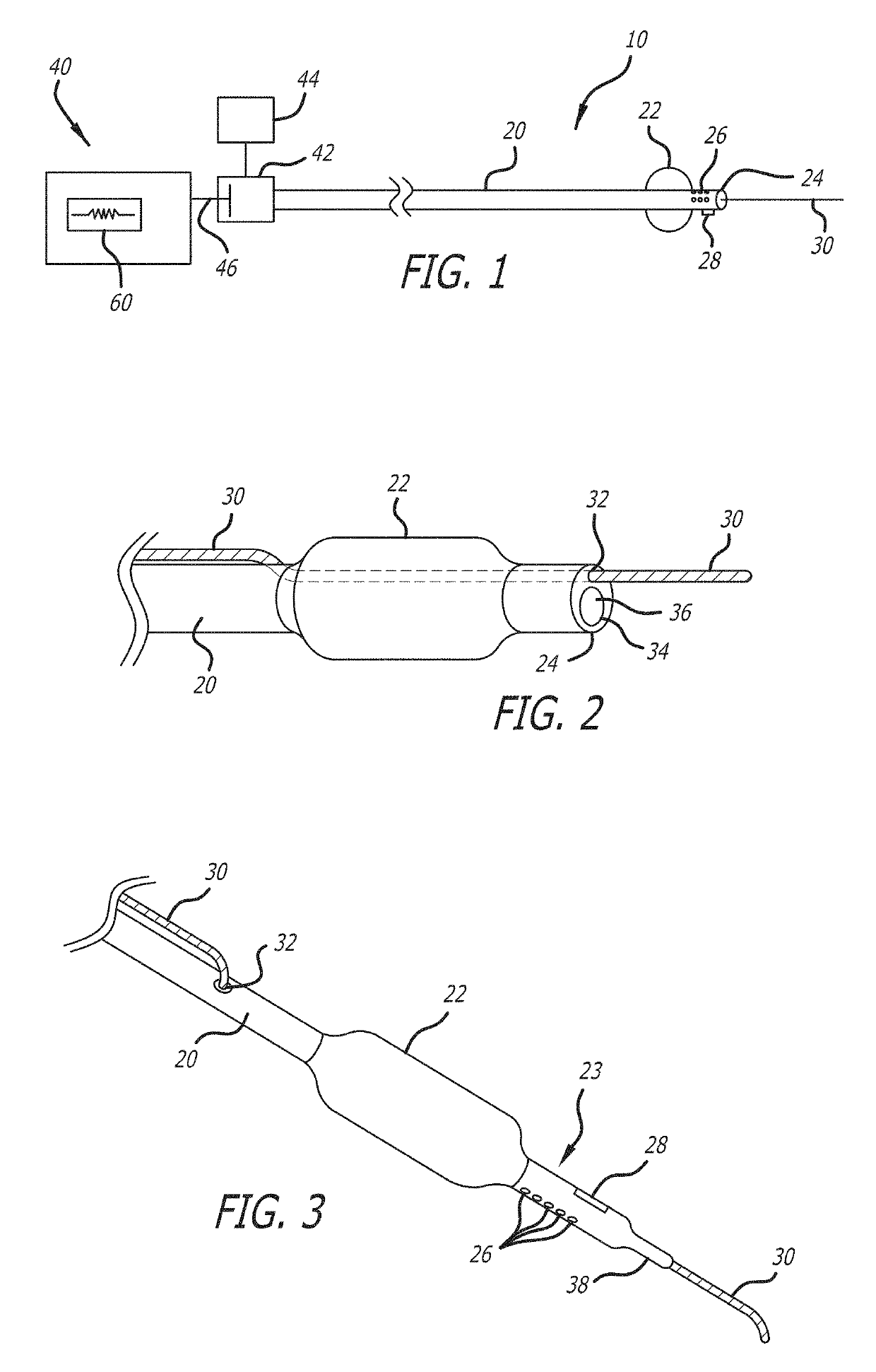
[0097]Referring now to the figures, and first to FIG. 1, there is shown an embodiment of a system 10 of the invention. The system 10 generally includes a catheter 20 with an occlusion balloon 22 near a distal end 24 of the catheter 20. The occlusion balloon 22 may be spaced apart from the distal end 24 enough to provide space for components occurring in some e...


PUM


Abstract

Microvascular obstruction is treated by introducing injectate into blood vessels affected by microvascular obstruction at precise flow rates, while blocking retrograde flow, such that the natural pumping of the heart aids in forcing the injectate into the affected microvessels. Monitoring pressure distal of an occlusion balloon is used to determine treatment effectiveness and heart health.

Description

RELATED APPLICATIONS[0001]This application claims priority to Provisional Patent Application Ser. No. 62 / 274,744, filed Jan. 4, 2016, entitled Method and Device for Alleviating Micro-Vascular Obstruction, Thrombus, or Platelet Obstruction in the Coronary Arteries, Heart or Other Tissues, and claims benefit of and priority to U.S. Provisional Application Ser. No. 62 / 320,230 filed Apr. 8, 2016 entitled Myocardial Pump, and claims benefit of and priority to U.S. Provisional Application Ser. No. 62 / 358,433 filed Jul. 5, 2016 entitled Myocardial Pump and Method, and claims benefit of and priority to U.S. Provisional Application Ser. No. 62 / 379,074 filed Aug. 24, 2016 entitled Myocardial Pump and Method, all of which are hereby incorporated herein by reference in their entireties.FIELD OF THE INVENTION[0002]Methods and devices for the diagnosis and treatment of microvascular obstruction (MVO) and other diseases of the microvasculature. More particularly, non-limiting embodiments include n...

Claims


Application Information

Patent Timeline
Patent Type & Authority Patents(United States)
IPC IPC(8): A61M25/10A61M5/172A61M5/145A61B17/22A61M5/142A61B90/00A61M25/00A61B17/3203
CPCA61M25/1011A61M5/142A61M5/1452A61M5/1723A61B17/22A61M2230/50A61B17/32037A61B2017/22054A61B2017/22067A61B2017/22071A61B2017/22084A61B2090/064A61M2005/14506A61M2025/0002A61M2205/502A61M2205/507A61M2230/04A61M2230/204A61B2017/00084A61B2017/22039A61B5/02007A61B5/0215A61B2017/00044
Inventor SCHWARTZ, ROBERT S.ROTHMAN, MARTIN T.HOEM, JON H.
Owner CORFLOW THERAPEUTICS AG
Learn More