Ceramidase and cell differentiation

a technology which is applied in the field of ceramidase and cell differentiation, can solve the problems of increasing the risk of transformation and/or contamination of the stem cell population, the current procedures are not very effective at maintaining the chondrogenic phenotype, and the risk of contamination, so as to improve the chondrogenic phenotype, improve cell quality, and reduce the number of apoptotic cells

Active Publication Date: 2019-07-16
MT SINAI SCHOOL OF MEDICINE
View PDF91 Cites 0 Cited by
  • Summary
  • Abstract
  • Description
  • Claims
  • Application Information

AI Technical Summary

Benefits of technology

[0009]A second aspect of the present invention relates to a method of improving the phenotype of a chondrocyte population. This method includes selecting a population of chondrocytes having a poor chondrocytic phenotype, and treating the selected cell population with a ceramidase to improve the chondrocytic phenotype of the population.
[0018]Ceramidases regulate the metabolism of several important bioactive lipids, including ceramide, sphingosine, and sphingosine-1-phosphate. The present invention shows that addition of recombinant rAC as a media supplement markedly improved the chondrogenic phenotype of expanded rat and horse articular chondrocytes, as well as human chondrocytes obtained from aged patients with osteoarthritis. The chondrocyte phenotype was assessed by a combination of histochemical staining (Alcian Blue and Safranin O), western blotting (e.g., Sox9, aggrecan, collagen IIA1, TGFβ1, FGF2, etc.), and / or qPCR. The effects of rAC were evident in monolayer and three-dimensional cultures, including cells grown on collagen scaffolds and fibrin gels. rAC also reduced the number of apoptotic cells in some culture conditions, contributing to overall improved cell quality. It is demonstrated herein that rat, equine, and human articular chondrocytes treated once with rAC in either monolayer cultures or three-dimensional scaffold systems had an improved chondrogenic phenotype after 2-3 week cell expansion, including enhanced expression of Sox9, FGF2, collagen II, aggregan and other chondrogenic markers.
[0019]In addition to these effects on primary chondrocytes, rAC had a significant impact on bone marrow-derived mesenchymal stem cells (MSCs). Here, it is shown that rat, feline, and equine bone marrow cells grown for only one week in the presence of rAC yield about two-fold more MSCs, and that rAC led to enhanced chondrogenesis of MSCs. When rAC was added to freshly harvested bone marrow cultures, an approximate two-fold enrichment of MSCs was observed by one-week. rAC also improved the chondrogenic differentiation of MSCs, as revealed by histochemical and immunostaining. These effects were observed using both rat and horse cells, and were synergistic with TGFβ1. These results demonstrate that rAC is an important supplement that could be used to improve the quality of cells used for cell-based cartilage repair, and can be useful in vivo to induce endogenous cartilage repair in combination with other techniques. Thus, rAC can be included in media used to culture primary chondrocytes and / or MSC chondrocyte differentiation media to improve the production and chondrogenic potential of these cells for cartilage repair. rAC also may added to three-dimensional scaffolds to induce and / or enhance chondrogenesis of cells in vivo. It is expected that other ceramidases may also be used for these purposes.

Problems solved by technology

An important limitation of this procedure, however, is the requirement of two invasive surgeries, the first of which requires extraction of cells from healthy cartilage tissue, and the second to implant the cells that have been expanded ex vivo.
Unfortunately, although many different culture systems have been used to evaluate chondrogenesis in vitro, no consensus method exists and current procedures are not very effective at maintaining the chondrogenic phenotype during the expansion period.
These procedures increase the risk of transformation and / or contamination of the stem cell population.

Method used


Image

  • Ceramidase and cell differentiation
  • Ceramidase and cell differentiation
  • Ceramidase and cell differentiation

Examples


example 1

and Methods

[0105]Animals—

[0106]Animals were raised under National Institutes of Health and USDA guidelines for the care and use of animals in research. Normal rats were derived from the MPS VI breeding colony (Kunieda et al., “Mucopolysaccharidosis Type VI in Rats: Isolation of cDNAs Encoding Arylsulfatase B, Chromosomal Localization of the Gene, and Identification of the Mutation,”Genomics 29(3):582-7 (1995), which is hereby incorporated by reference in its entirety) maintained at the Mount Sinai School of Medicine, while the larger animals (cats and horses) were maintained at the University of Pennsylvania School of Veterinary Medicine. The animals were housed with ad libitum food and water. Euthanasia was performed on cats using 80 mg / kg of sodium pentobarbital (Veterinary Laboratories, Lenexa, Kans.) in accordance with the American Veterinary Medical Association guidelines. Euthanasia of rats was performed using carbon dioxide inhalation. Equine cells were obtained during routin...

example 2

n Sphingolipid Metabolism Following rAC Treatment of Primary Articular Chondrocytes

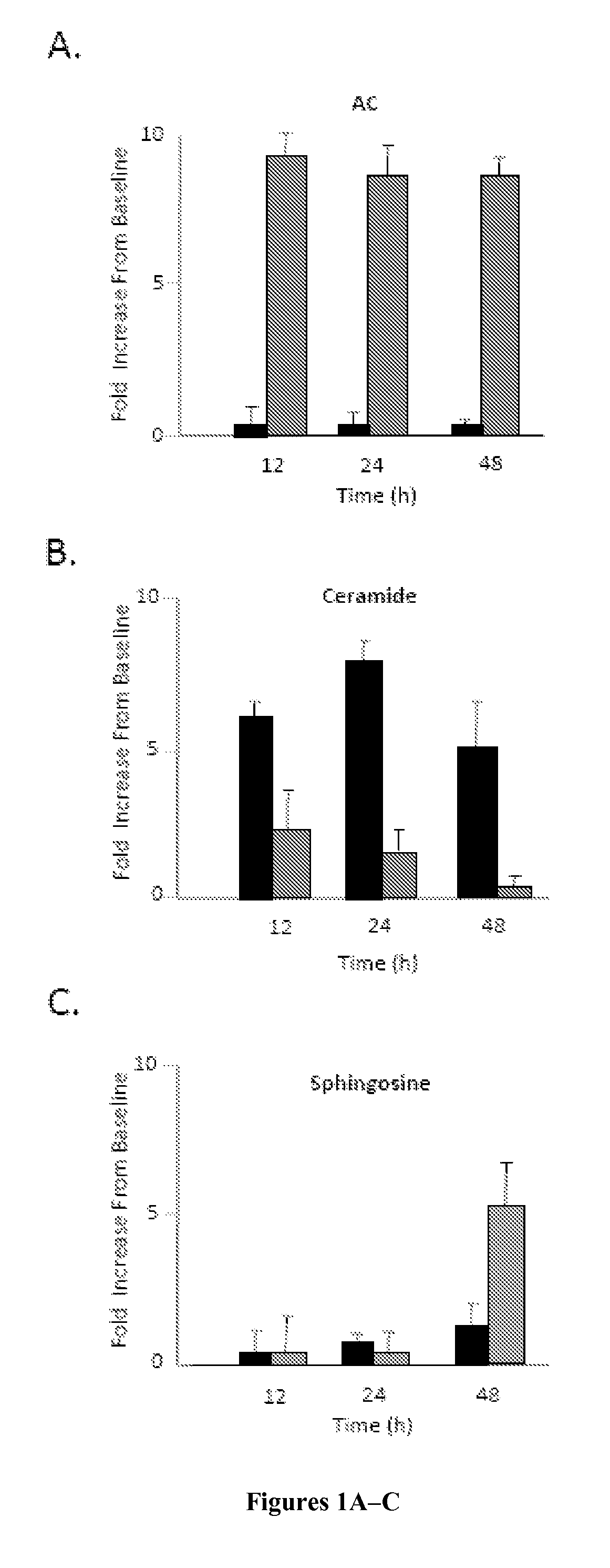
[0136]Primary rat articular chondrocytes were isolated and grown in monolayer cultures as described in Example 1 using DMEM containing 10% FBS with or without rAC. For the baseline timepoint, cells were collected from 6 rats and pooled without culture. The acid ceramidase, ceramide, and sphingsoine levels in these cell lysates were compared to cells grown for 12, 24, and 48 hours with or without rAC. The data is summarized in FIGS. 1A-1C.

[0137]As expected, cells grown in media supplemented with rAC exhibited markedly increased AC activity by 12 hours, and this was sustained through 48 hours. Ceramide levels were also increased compared to baseline by 12 hours, but less in cells exposed to rAC. In fact, by 48 hours the ceramide levels in the rAC-treated cells had returned to baseline, while in those without rAC treatment remained significantly elevated. Also, by 48 hours significant elevations of sphin...

example 3

f rAC Supplementation on Expanded Monolayer Cultures of Primary Articular Chondrocytes

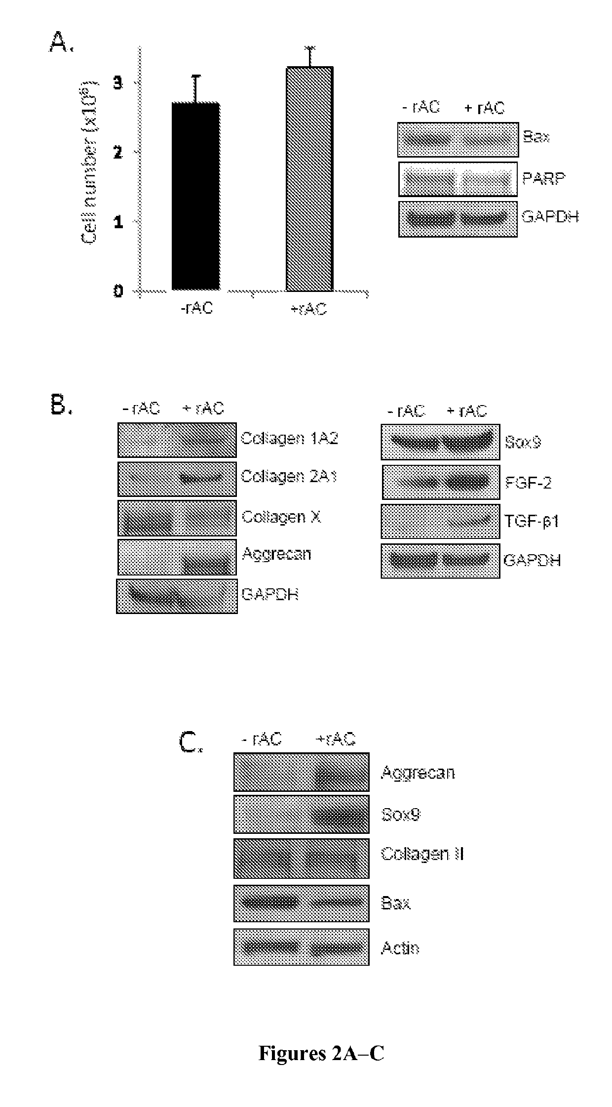
[0139]The effects of rAC supplementation on the phenotype of articular chondrocytes after cell expansion were next evaluated. For these experiments, rat chondrocytes were grown for three weeks with and without rAC in DMEM containing 10% FBS. rAC was added once at the time of the initial cell plating (P0). All analyses were performed at the end of the three-week expansion period unless otherwise mentioned.

[0140]No effect of rAC treatment on the total number of cells was observed (FIG. 2A), consistent with the fact that the levels of two apoptotic markers, Bax and PARP, were also very similar with and without rAC treatment.

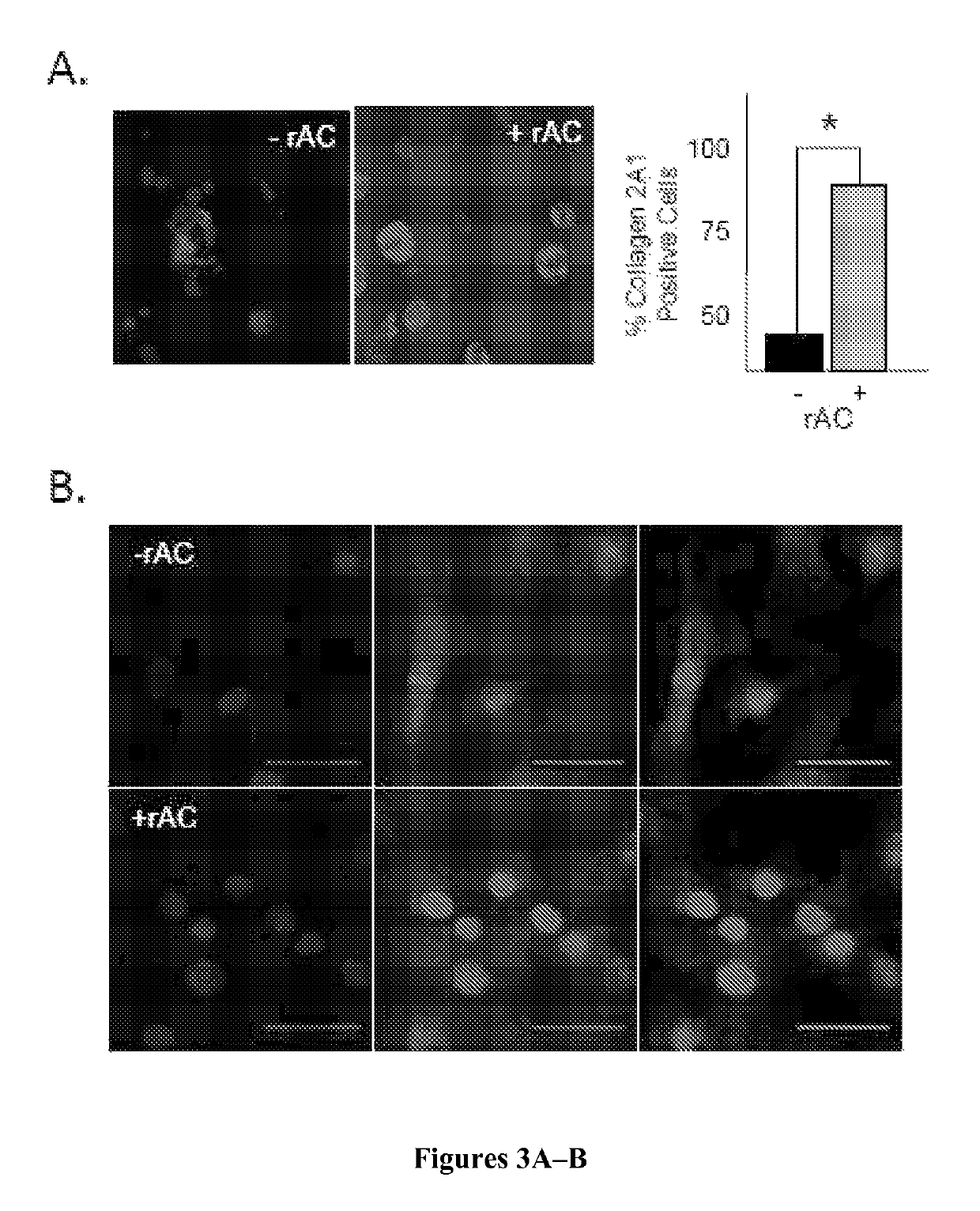
[0141]However, despite obtaining the same number of cells, there was a marked effect of rAC treatment on cell quality at the end of the 3-week expansion period, as determined by the expression of various chondrocyte markers. As shown in FIG. 2B, western blot analysis revealed that ...


PUM

PropertyMeasurementUnit
v/vaaaaaaaaaa
v/vaaaaaaaaaa
v/vaaaaaaaaaa

Abstract

The present invention relates to methods of producing chondrocytes, improving the phenotype of a chondrocyte population, promoting chondrogenesis, maintaining a cell population in a differentiated state or increasing the number of cells of a population in a differentiated state, producing a population of differentiated cells, and treating certain diseases or disorders. Each of these methods involves treating a cell population with, and / or culturing a cell population in, a ceramidase.

Description

[0001]This application is a national stage application under 35 U.S.C. § 371 of PCT Application No. PCT / US2012 / 054316, filed Sep. 7, 2012, which claims the benefit of U.S. Provisional Patent Application Ser. No. 61 / 531,917, filed Sep. 7, 2011, all of which are hereby incorporated by reference in their entirety.FIELD OF THE INVENTION[0002]This invention relates to ceramidase and cell differentiation.BACKGROUND OF THE INVENTION[0003]Cell-based therapy for cartilage repair has gained increasing popularity since the first reports of successful autologous chondrocyte implantation (ACI) over 10 years ago (Minas et al., “Chondrocyte Implantation in the Repair of Chondral Lesions of the Knee: Economics and Quality of Life,”Am. J. Orthop. 27(11):739-44 (1998)). In ACI, primary chondrocytes are obtained from small biopsies of healthy articular cartilage, expanded, and then placed onto three-dimensional scaffolds for subsequent use in cartilage repair surgery (see Vavken et al., “Effectiveness...

Claims


Application Information

Patent Timeline
Patent Type & Authority Patents(United States)
IPC IPC(8): C12N5/077A61K38/50A61K35/32A61K38/18
CPCA61K38/50A61K35/32A61K38/1841C12N5/0655C12Y305/01023C12N2506/1353C12N2501/73A61K2300/00A61P19/00A61P21/00A61P25/00A61P3/00
Inventor SCHUCHMAN, EDWARD H.SIMONARO, CALOGERA M.
Owner MT SINAI SCHOOL OF MEDICINE
Why Eureka
Try Eureka