
Method of analysing DNA sequences

a dna sequence and sequence technology, applied in the field of dna sequence analysis, can solve the problems of not readily providing quantitative estimates of megabase-scale chromosomal interactions, sensitivity possible with this previous design, and high cost per sample, so as to reduce the concentration of oligo pools and increase the efficiency of double capture

Active Publication Date: 2021-03-02
OXFORD UNIV INNOVATION LTD
View PDF14 Cites 0 Cited by
  • Summary
  • Abstract
  • Description
  • Claims
  • Application Information

AI Technical Summary

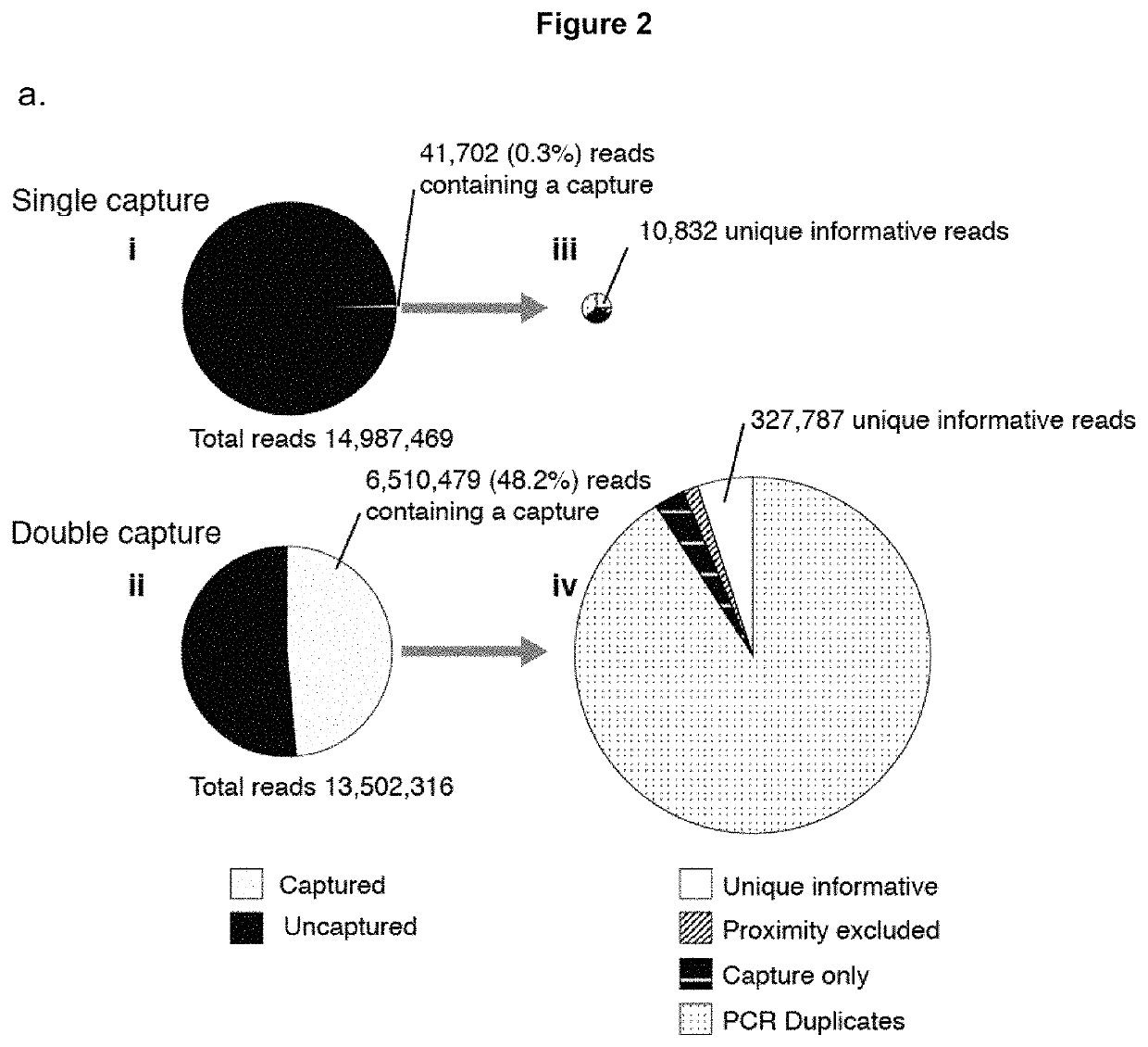
Benefits of technology

[0010]A further adaptation of the original Capture-C method is the Capture-Hi-C method (as exemplified in WO2015 / 033134) which combines the Capture-C method with the Hi-C library production. The Capture-Hi-C method requires the superimposition of a biotin group during the ligation step of library production to capture (as illustrated in FIG. 1 of WO2015 / 033134). This step is inefficient and so can greatly decrease the complexity of the library; ultimately, it greatly limits the sensitivity of the approach. Loss of complexity in the library is directly related to the amount of information that can be extracted per cell of the original sample. Therefore methods which involve such losses are only readily usable in circumstances where very large numbers of cells are available.
[0012]Analysis of the original Capture-C data showed that the sonication step allowed the identification of PCR duplicates in this assay, an effect that is problematic in most existing 3C methods. This allowed for the direct measurement of the efficiency of enrichment for ligation junction in the assay and made it possible to determine when all information had been extracted from a given library and where further sequencing would not yield any further information. By performing an experiment targeting a single gene promoter, it was surprisingly discovered that the captured DNA from this single region of interest made up less than 1% of the sequenced reads. This showed that great sequencing depth would be required in order to extract all of the information from the library with standard single capture. This would be completely impractical for smaller designs or repeated experiments, and it showed that simplifying the design itself would not readily increase the signal to noise ratio. It suggested that the signal to noise ratio was in fact inherent to the then current capture protocol.
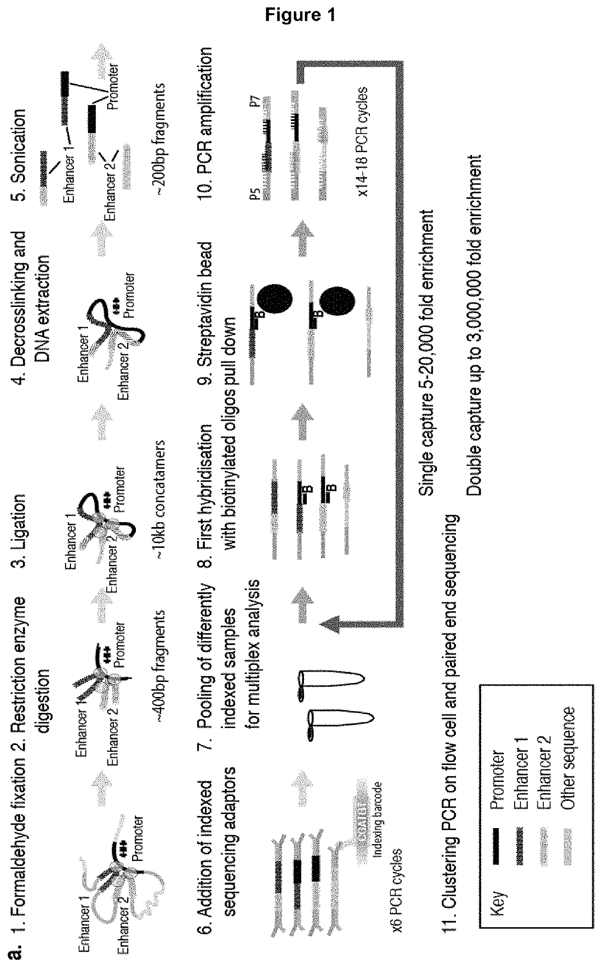
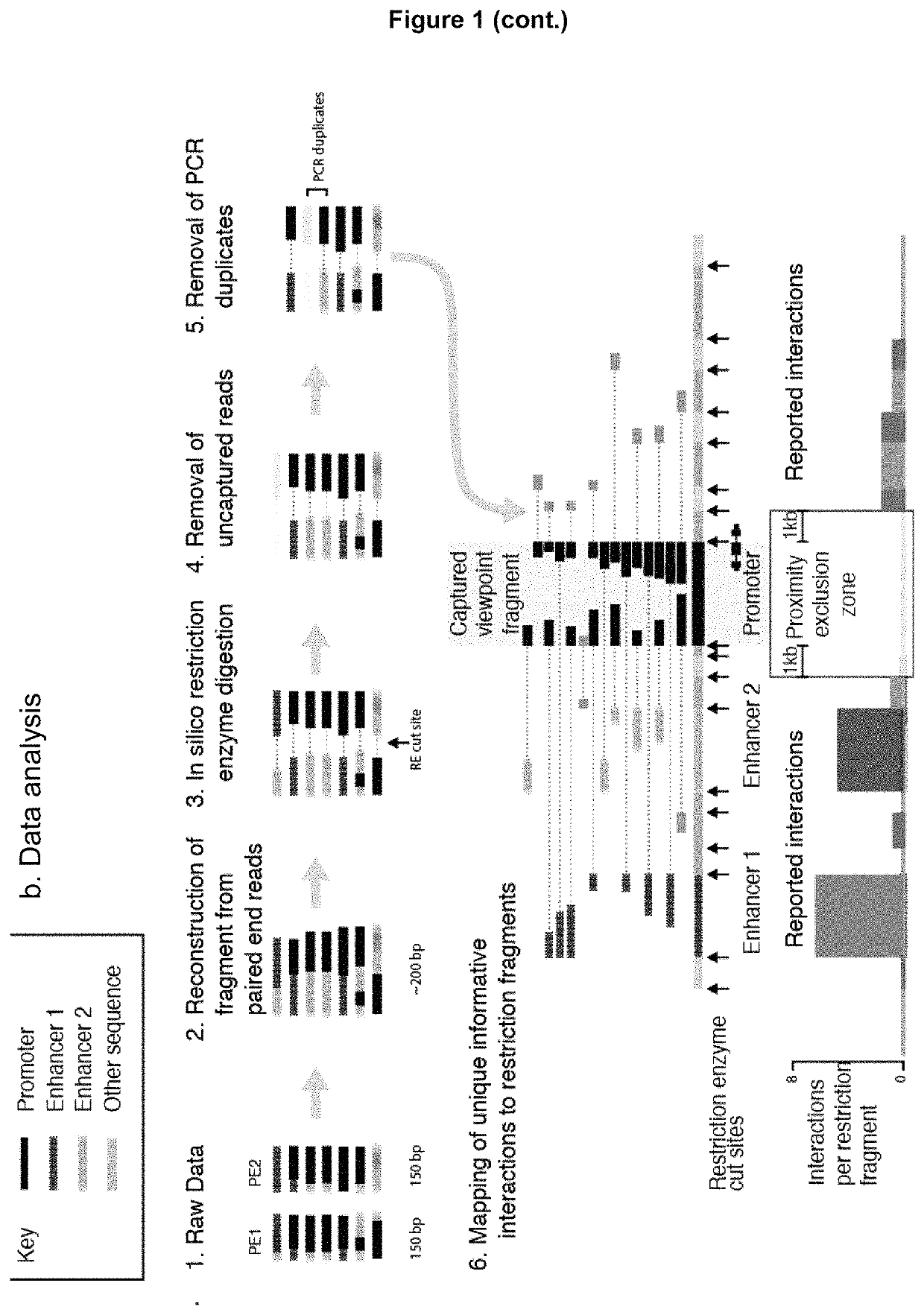
[0015]Due to the complete control over PCR duplicates in the Capture-C protocol, it was realised that the library could be substantially over-amplified in the initial step of the library preparation part of the protocol, so that each informative junction would be represented multiple times prior to capture. It was further realised that, as junctions were now robustly represented and that the background was not intrinsic to the capture probes themselves, then the remaining background could substantially further mechanically-depleted by a second round of capture without loss of complexity.
[0016]The inventors have now found that the use of two sequential oligonucleotide capture steps applied to a 3C library (e.g. a PCR duplicated 3C library) prior to sequencing results in up to 3,000,000 fold enrichment compared to an uncaptured 3C library so that captured material now makes up approximately 50% (rather than 1%) of the sequenced material. This second capture step increases the number of PCR cycles and the number of PCR duplicates sequenced because the library complexity (i.e. the number of interactions available to capture) limits the number of unique interactions that could be sequenced. The greatly improved enrichment means that the depth of sequencing is no longer limiting. Using this new method, any PCR duplicates can be easily and efficiently excluded bioinformatically.
[0017]Furthermore, due to the huge increase in signal, independent 3C libraries (e.g. from different cell types or different stages of development) can now be captured and processed in a single tube making separately-indexed samples directly comparable. This greatly increases throughput and allows meaningful subtractive analysis of chromosome conformation in different cell types.
[0020]The investigation of gene regulation is not only limited by the number of genes or elements that can be interrogated, but also by the number of replicates, conditions, cell types and genetic variants that can be easily analysed. The huge increase in signal of NG Capture-C allows for the simultaneous capture of multiple samples in a single reaction, greatly increasing the throughput and economy of the assay. In practice, this allows complete networks of important genes, such as those encoding the Yamanaka pluripotency factors [32] (Myc, Sox2, Oct4, Klf4) to be analysed simultaneously in multiple cell types. The data are compatible with standard analytical tools and their reproducibility and comparability between active and inactive states of NG Capture-C provides a complementary approach to the statistical identification of regulatory elements. This complementary approach is capable of identifying all known regulatory elements at well characterised test loci, at levels of resolution previously not possible. Importantly, mindful of the current challenges in the analysis of GWAS and regulatory variants, the NG Capture-C method can been optimized to be effective at smaller cell numbers (approximately 100,000 cells) and to generate SNP-specific interaction profiles.

Problems solved by technology

The sheer variety of techniques which are currently available creates a challenge when attempting to improve yet further on the sensitivity of the basic 3C technique.
All 3C techniques require an enrichment step, the basis of which varies across most of the methods and it often unclear which will prove to be the most efficient and flexible approach.
Furthermore, issues such as the choice of restriction endonucleases or fragmentation method, cross-linking stringency, primer design, library complexity and probe position can all have an effect on the efficacy of any one particular enrichment method.
The original Capture-C protocol [12] used oligos synthesized on a microarray (Agilent SureSelect) with a minimum design of 40,000 oligos, irrespective of the number of desired viewpoints; the cost per sample was therefore very high for small designs.
Furthermore, the sensitivity possible with this previous design did not readily allow for the analysis of very long-range cis-interactions or trans-interactions and did not provide quantitative estimates of megabase-scale chromosomal interactions.
Importantly, although the method was high throughput at the level of which regions of the genome it could analyse simultaneously, it was still limited to one sample per assay.
This step is inefficient and so can greatly decrease the complexity of the library; ultimately, it greatly limits the sensitivity of the approach.
Loss of complexity in the library is directly related to the amount of information that can be extracted per cell of the original sample.
Therefore methods which involve such losses are only readily usable in circumstances where very large numbers of cells are available.

Method used


Image

  • Method of analysing DNA sequences
  • Method of analysing DNA sequences
  • Method of analysing DNA sequences

Examples


example 1

on of 3C Libraries

[0124]Single cell preparations of erythroid cells were made by gently dissociating cells from the spleen of a mouse treated with phenylhydrazine (40 mg / g body weight×3 doses 12 h apart; sacrificed on day 5). Phenylhydrazine causes haemolytic anemia and marked erythroid expansion in the spleen so that 80% or more of cells are erythroid cells (as defined by CD71+ ter119+). The cells were passed through a 40 μm cell strainer to remove clumps. For ter119 selection, cells were stained with ter119-phycoerythrin (PE) and purified using anti-PE MACS beads (Miltenyi Biotec) prior to fixation with formaldehyde. Mouse E14 ES cells were trypsinised and washed once prior to fixation.

[0125]Each aliquot of 107 cells was resuspended in 10 ml of RPMI with 10% FCS in a 15-ml conical centrifuge tube. 549 μl 37% (vol / vol) formaldehyde was added to each aliquot to make an overall concentration of 2% (vol / vol). A 10 minute incubation was performed at room temperature on a roller mixer. ...

example 2

of Sequencing Adaptors

[0132]5 μg of 3C library was sonicated to 200 bp using a Covaris S220 Focussed ultrasonicator (6 cycles of 60 s: duty cycle 10%; intensity 5; cycles per burst 200). The degree of sonication was confirmed using an Agilent Bioanalyser or Tapestation (DNA 1000). Illumina Truseq indexed sequencing adapters were added using NEBnext reagents (E6000 / E6040 / E7335 / E7500). This involved end repair, addition of overhanging A bases, ligation of adapters and PCR to add the indices. The DNA was cleaned up between reactions using Ampure XP beads at a 1:1.8 ratio for all clean up steps to minimize the selection of larger fragments and losses of material were minimised. 6-8 cycles of PCR were used when addition the Truseq indices using the Agilent Herculase II PCR kit. Generally 1.5-2 μg of adapter ligated material was generated, however, to maximize library complexity the library preparation was usually done in duplicate (to use 10 μg of input material) and the samples were poo...

example 3

eotide Capture

[0134]1.5-2 μg of adapter ligated material was placed in a 1.5 ml microcentrifuge tube with 5 μg COT DNA from the appropriate species; 1000 pM Nimblegen HE Universal blocking oligo and 1000 pM Nimblegen HE Index specific blocking oligo (corresponding to the Illumina TS index used). The sample was then dried using a vacuum centrifuge (50-60° C.) until no liquid remained. The residue was dissolved in 7.5 μl Nimblegen Hybridization Buffer and 3 μl Nimblegen Hybridization Component A followed by denaturation at 95° C. for 10 minutes. Concurrently 4.5 μl of the biotinylated capture oligonucleotide library (total 13 pM) was heated to in a 0.2 ml PCR tube to 47° C. in a PCR block. After 10 minutes the 3C library and blocking oligonucleotides were added to the preheated biotinylated oligonucleotides at 47° C. The hybridization reaction was incubated in a PCR machine at 47° C. for 64-72 h (with a heated lid at 57° C.).

[0135]The Nimblegen SeqCap EZ Wash Buffers (I, II, III, Stri...


PUM

PropertyMeasurementUnit
volumeaaaaaaaaaa
volumeaaaaaaaaaa
volumeaaaaaaaaaa

Abstract

The present invention relates to a method of identifying nucleic acid regions within a nucleic acid sample which interact with one another. In particular, the method relates to a chromatin conformation capture (3C) method which may be used to analyse the interactions between enhancers, silencers, boundary elements and promoters at individual loci at high resolution.

Description

CROSS-REFERENCE[0001]This application is a section 371 U.S. National phase of PCT / GB2016 / 053314, filed Oct. 24, 2016 which claims priority from GB 1518843.6, filed Oct. 23, 2015, which is incorporated by reference in its entirety.FIELD OF THE INVENTION[0002]The present invention relates to a method of identifying nucleic acid regions within a nucleic acid sample which interact with one another. In particular, the method relates to a chromatin conformation capture (3C) method which may be used to analyse the interactions between enhancers, silencers, boundary elements and promoters at individual loci at high resolution.BACKGROUND OF THE INVENTION[0003]Progress in our ability to annotate regulatory elements in the genome and determine their potential function has been driven by technological advances, such as RNA-seq [1], ChIP-seq [2, 3], DNase-seq [4] and ATAC-seq [5]. However, an outstanding challenge is to understand the mechanisms by which regulatory elements control specific gene...

Claims


Application Information

Patent Timeline
Patent Type & Authority Patents(United States)
IPC IPC(8): C12Q1/6827
CPCC12Q1/6827C12Q2600/156C12Q2523/101C12Q2535/122C12Q2563/131
Inventor HUGHES, JAMES R.DAVIES, JAMES
Owner OXFORD UNIV INNOVATION LTD
Learn More