
Nanocapsule delivery system for ribonucleoproteins

a ribonucleoprotein and nanocapsule technology, applied in the direction of viruses/bacteriophages, peptide/protein ingredients, genetic material ingredients, etc., can solve the problems of delivering rnps using nonviral biomaterials specifically, plasmids carrying the risk of insertional mutagenesis into the host genome, and delivering crispr nuclease along with its single-guide rna (sgrna) into cells,

Active Publication Date: 2021-05-25
WISCONSIN ALUMNI RES FOUND
View PDF3 Cites 0 Cited by
  • Summary
  • Abstract
  • Description
  • Claims
  • Application Information

AI Technical Summary

Benefits of technology

[0007]While not wishing to be bound by theory, it is believed that the polymer shell protects the RNP from degradation in the extracellular environment and assists in delivery of the RNP into cells as shown in the schematic of FIG. 1B. The small size of the nanocapsules (e.g., having a hydrodynamic diameter of about 7 to about 25 nm) as well as any CPP and / or targeting ligand present allow the nanocapsules to enter the target cells by endocytosis. The presence of the imidazolyl groups in the polymer shell facilitate endosomal escape of the nanocapsules, because imidazole groups, having a pKa value of ˜6.0, can take up protons in the acidic endosomes. This absorption of protons leads to endosomal-membrane disruption as previously described (G. Chen, Y. Wang, R. Xie, S. Gong, J. Control. Release 2017, 259, 10), thereby enabling release of the nanocapsules into the cytosol. There, due to the glutathione (GSH)-rich environment of the cytosol (2-10 mM), the disulfide bonds of the biodegradable cross-linkers are reduced, causing the polymer shell to degrade. Once released from the nanocapsules, RNPs can enter the nucleus, optionally facilitated by one or more nuclear localization signals (NLSs), on Cas9 proteins, and perform its gene editing functions.

Problems solved by technology

For somatic gene editing applications both in vivo and ex vivo, delivery of the CRISPR nuclease along with its single-guide RNA (sgRNA) into cells is a significant challenge.
Viral delivery of CRISPR components is commonly used for delivery of the nuclease and sgRNA into cells, but viral vectors raise additional safety concerns for clinical application.
Plasmids also carry the risk of insertional mutagenesis into the host genome.
To date, however, delivering RNPs using nonviral biomaterials specifically to a cell type of interest within the body or ex vivo in culture occurs with only limited success.

Method used


Image

  • Nanocapsule delivery system for ribonucleoproteins
  • Nanocapsule delivery system for ribonucleoproteins
  • Nanocapsule delivery system for ribonucleoproteins

Examples


example 1

on of RNP

[0096]sgRNAs In Vitro Transcription. A DNA double stranded template of a truncated T7 promoter and desired sgRNA sequence was formed through overlap PCR using Q5 high fidelity polymerase (New England Biolabs) according to manufacturer's protocols and was placed in the thermocycler for 35 cycles of 98° C. for 5 s, 52° C. for 10 s, and 72° C. for 15 s, with a final extension period of 72° C. for 10 min. PCR products were then incubated overnight at 37° C. in a HiScribe T7 in vitro transcription reaction (New England Biolabs) according to manufacturer's protocol. The resulting RNA was purified using a MEGAclear Transcription Clean-Up Kit (Thermo Fisher). sgRNA concentration was quantified using a Qubit fluorometer (Thermo Fisher). The sgRNA sequence targeting mCherry (with PAM site) is as follows: GGAGCCGTACATGAACTGAG GGG (SEQ ID NO: 46).

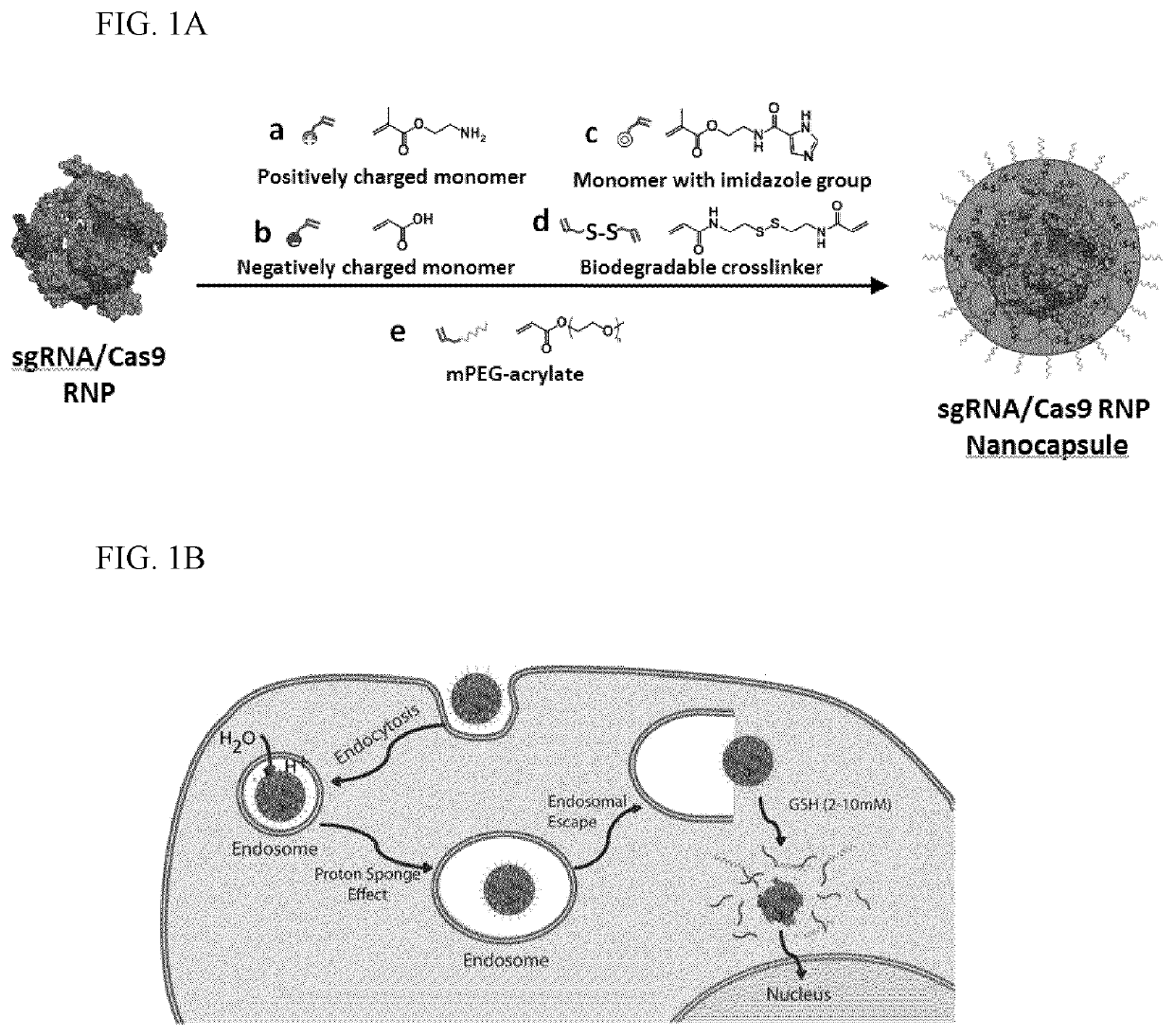
[0097]Preparation of Nanocapsules (NCs), Method I. sNLS-Cas9-sNLS protein (Aldevron, Madison, Wis.) was combined with the desired sgRNA at a ...

example 2

ization of RNP NCs

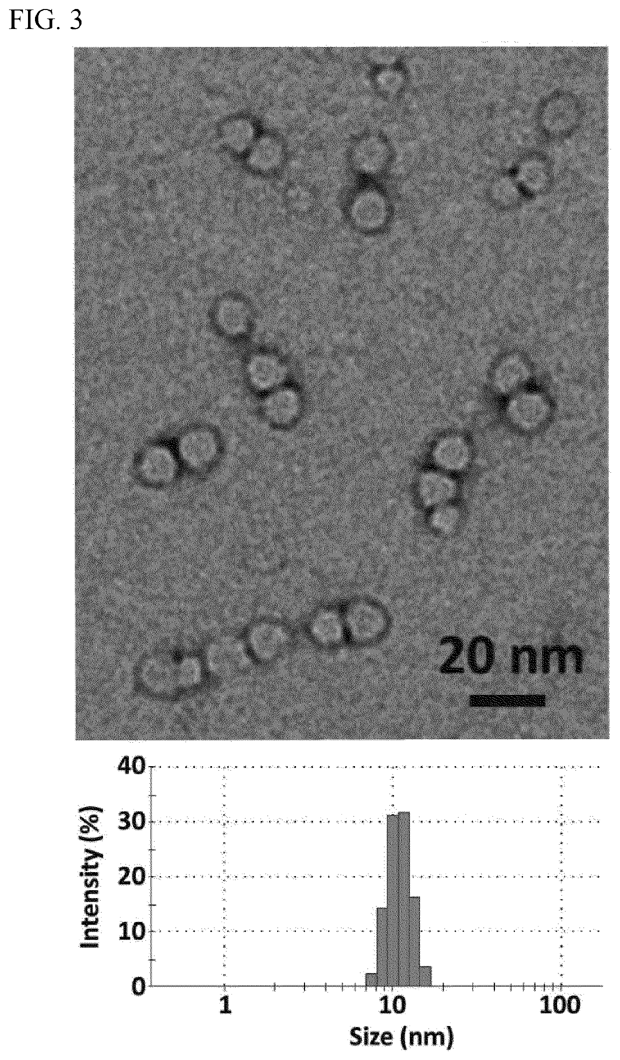
[0100]The sizes and morphologies of RNP NCs were studied by dynamic light scattering (DLS, ZetaSizer Nano ZS90, Malvern Instruments, USA) and transmission electron microscopy (TEM, FEI Tecnai G2 F30 TWIN 300 KV, E.A. Fischione Instruments, Inc. USA). Zeta potentials were measured by ZetaSizer Nano ZS90 (Malvern Instruments, USA). After purification, transmission electron microscopy (TEM) of dried NC-3 indicated uniformly sized NCs with an average size of ˜10 nm (FIG. 3). The average hydrodynamic diameter measured by DLS was 12.7 nm and the zeta potential of −0.9 mV was obtained for NC-3 (Table 1), indicating that the net negative charge of the RNP masked by the NC.

[0101]

TABLE 1Average hydrodynamic diameter and zeta potential of Cas9,RNP, and RNP NC (prepared by Method I).Cas9 proteinRNPRNP NC-3Size (nm)6.3 ± 0.6   7.6 ± 1.1 12.7 ± 2.8Zeta potential (mV)7.9 ± 3.13−18.5 ± 4.1−0.9 ± 1.3

example 3

RNP Delivery and Efficacy

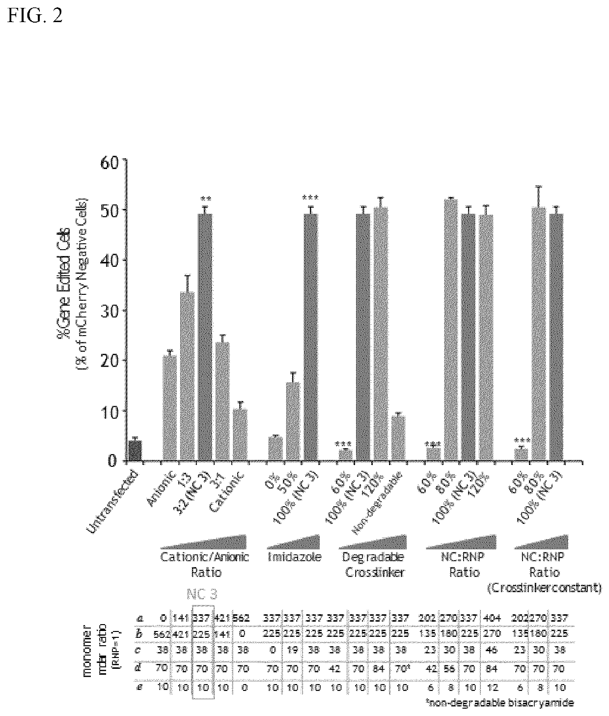
[0102]Cells were seeded in 96 well plates (Table 2) in 100 μL of media one day prior to transfection. On the day of transfection, RNPs with the desired guide sequence were formed, NC-RNP or Lipofectamine 2000-RNP (0.75 μL Lipofectamine 2000 / well) complexes were added to the cells for transfection of RNPs. At day 6, cells were collected for flow cytometry (BD FACSCanto II) and assayed for mCherry fluorescence. Flow analysis was performed using FlowJo software.

[0103]

TABLE 2Conditions for in vitro functional testing of nanocapsules (NCs).Seeded cellnumber perPlateCas9 doseCell Typewelltreatment(ng / well)Human50000.1% Gelatin125EmbryonicKidney (HEK)Human6500Matrigel350embryonic stemcells (WA09)Jurkats5000N / A350M21 Melanoma50000.1% Gelatin250NHDF50000.1% Gelatin250Fibroblasts

[0104]Genomic analysis. DNA was isolated from cells using DNA QuickExtract (Epicentre, Madison, Wis.) following treatment by 0.05% trypsin-EDTA and centrifugation. QuickExtract solution was in...


PUM

PropertyMeasurementUnit
mass ratioaaaaaaaaaa
weight average molecular weightaaaaaaaaaa

Abstract

Provided herein are nanocapsules comprising a single ribonucleoprotein (RNP) complex as a core and an biodegradable crosslinked polymer shell that encapsulates the core, wherein the RNP complex comprises a Cas9 polypeptide and a guide RNA, and the biodegradable crosslinked polymer shell comprises polymerized monomers of imidazolyl acryloyl monomers, bisacryloyl disulfide monomers (a biodegradable cross-linker), optionally PEG acryloyl monomers, and either cationic acryloyl monomers, anionic acryloyl monomers, or both cationic and anionic acryloyl monomers (optionally in combination with non-ionic acryloyl monomers) as defined herein. Also provided are methods of making the nanocapsules, kits containing the nanocapsules and methods of delivering the encapsulated RNP to cells.

Description

CROSS-REFERENCE TO RELATED APPLICATIONS[0001]This application claims the benefit of and priority to U.S. Provisional Patent Application No. 62 / 551,202, filed Aug. 28, 2017, the entire contents of which are incorporated herein by reference.GOVERNMENT RIGHTS[0002]This invention was made with government support under GM119644 and awarded by the National Institutes of Health. The government has certain rights in the invention.SEQUENCE LISTING[0003]The instant application contains a Sequence Listing which has been filed electronically in ASCII format and is hereby incorporated by reference in its entirety. Said ASCII copy, created on Oct. 24, 2018, is named 032026-1379_SL.txt and is 14,508 bytes in size.FIELD[0004]The present technology relates generally to the field of encapsulated ribonucleoprotein delivery systems. The compositions of such systems include a polymeric shell encapsulating a Cas9 polypeptide complexed with a guide RNA.BACKGROUND[0005]Gene editing is anticipated to modify...

Claims


Application Information

Patent Timeline
Patent Type & Authority Patents(United States)
IPC IPC(8): A61K9/51A61K48/00A61K47/32C12N15/11A61K38/46C12N15/113A61K31/7105A61K9/50C12N9/22C12N15/90
CPCA61K9/51A61K9/5026A61K9/5138A61K31/7105A61K38/46A61K47/32A61K48/0041A61K48/0091C12N9/22C12N15/11C12N15/111C12N15/113C12N15/907C12N2310/20C12N2320/32C12N2800/80
Inventor GONG, SHAOQINABDEEN, AMRSAHA, KRISHANUCHEN, GUOJUNWANG, YUYUANXIE, RUOSEN
Owner WISCONSIN ALUMNI RES FOUND
Learn More