Bacterial membrane nanoparticles as an immunotherapy system for cancer treatment

Active Publication Date: 2020-03-12
WISCONSIN ALUMNI RES FOUND
View PDF1 Cites 0 Cited by
  • Summary
  • Abstract
  • Description
  • Claims
  • Application Information

AI Technical Summary

Benefits of technology

[0006]While not wishing to be bound by theory, FIGS. 1A and 1B show an illustrative embodiment of a method of use of the present nanoparticles in which antigen capture groups are present on the surface of the nanoparticles. In this embodiment, BNP is intratumorally injected following radiation therapy (RT) treatment (FIG. 1A). The BNP captures exposed cancer necantigens through antigen capture groups such as maleimide groups. The BNP undergoes highly efficient uptake in antigen-presenting cell (APC) and stimulates immune response due to the presence of its bacterial membrane component shell. The TLR2 and TLR4 agonists among the bacterial membrane components help stimulate the innate immune response and the eventual development of the adaptive immune response. Once the BNP is endocytosed (FIG. 1B), the anionic immune adjuvant is released from the polyplex core and is believed to activate toll-like receptor 9 (TLR-9), which is located at the endosomal inner membrane, thus prompting the maturation of the APC. Tran

Problems solved by technology

Transferring the neoantigen from the endosome into the cyt

Method used


Image

  • Bacterial membrane nanoparticles as an immunotherapy system for cancer treatment
  • Bacterial membrane nanoparticles as an immunotherapy system for cancer treatment
  • Bacterial membrane nanoparticles as an immunotherapy system for cancer treatment

Examples


Example

Example 1. Synthesis of PC7A Polymer

[0043]Synthesis of Methacrylate Monomer.

[0044]The monomer 2-(hexamethyleneimino) ethyl methacrylate (C7A-MA) was synthesized according to previous reports (K. J. Zhou, Y. G. Wang. X. N. Huang, K. Luby-Phelps, B. D. Sumer, J. M. Gao, Angew Chem Int Edit 2011, 50, 6109). Briefly, 1H-azepine-1-ethanol, hexahydro- (5.0 g, 34.5 mmol) and triethylamine (TEA, 7.0 g, 69 mmol) was dissolved in 100 mL dried THF and cooled to 0° C. in an ice bath. Methacryloyl chloride (4.0 g, 38.5 mmol) was dissolved in 15 mL dried THF and slowly dropped into the previous solution. The reaction was moved to room temperature and stirred for 8 h, then filtered to remove TEA salt. The filtrate was condensed by rotary evaporation and further purified by flash chromatography (Hex:EtOAc=2:1). C7A-MA: 1H NMR (TMS, CDCl3, ppm): 6.1 (s, 111), 5.6 (s, 1H), 4.2 (t, 2H), 2.8 (s, 2H), 2.7 (s, 4H), 2.0 (s, 3H), 1.8 (s, 4H), and 1.6 (s, 4H).

[0045]The monomer 2-(tert-butoxycarbonylamino) e...

Example

Example 2. Bacteria Growth and Membrane Extraction

[0052]M. smegmatis (strain mc2 155) was grown in Middlebrook 7H9 broth with the addition of a 10% (vol / vol) FD019 supplement at 37° C. with aeration and shaking. Cells were harvested at an OD600 of around 3, collected with a 4000 G centrifuge, and washed twice with distilled water.

[0053]The bacterial membrane was extracted as follow. A ˜2 g bacteria pellet was dispersed in 40 mL Tris HCl buffer (20 mM, pH 8.0) containing 15 mg / mL lysozyme. It was incubated in a 37° C. shaker for 3 h. After adding 400 mg SDS, the cells were further broken down with a probe sonicator in an ice bath for 10 min and then lyophilized. The dried bacteria lysate was suspended in a chloroform-methanol-water solution (CMW; 30:15:1, v / v / v), placed on a shaker at 37° C. for 1 h, and then filtered by Millipore HVLP Durapore® membrane (0.45 gun). The residue was extracted by CMW two more times and then the filtrate was combined and dried under vacuum. The bacteria...

Example

Example 3. Preparation of the BNP

[0054]Preparation and characterization of PC7A / CpG polypex.

[0055]Polyplex was freshly prepared by adding the polymer solution at different concentrations (1:1, 2.5:1, 5:1, 10:1, 20:1) into 1.2 mg / mL CpG solution (1:1, v / v). The mixture was vortexed for 10 seconds and incubated at room temperature for 15 min before use. Agarose gel electrophoresis was employed to optimize the PC7A to CpG weight ratios at different pHs (6.5, 7.4) using a 1% agarose gel in a TAE (Tris-acetate-EDTA) buffer solution with a current of 110 V for 30 min. The retardation of the complexes was stained by Sybr safe and visualized on a UV illuminator (Bio-Rad Baloratories, Inc., Hercules, Calif., USA). Based on the electrophoresis results (data not shown), CpG was completely complexed at a pH of 7.4 when the polymer / CpG weight ratio reached 2.5 and above.

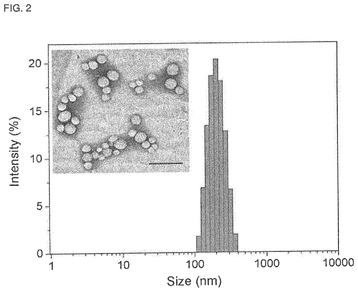
[0056]Preparation and Characterization of BNP.

[0057]Seven microliters of a 1.5M sodium carbonate solution and 100 μL freshly pr...


PUM

PropertyMeasurementUnit
Hydrodynamic diameteraaaaaaaaaa
Weight ratioaaaaaaaaaa
Responsivityaaaaaaaaaa

Abstract

Provided herein are nanoparticles comprising a polyplex core comprising one or more pH-responsive polymers and one or more anionic immune adjuvants, wherein each pH-responsive polymer comprises ionizable amine groups; and a shell of bacterial cell membrane components at least partially coating the polyplex core, wherein the bacterial cell membrane components comprise TLR 2 and/or TLR 4 agonists. Also provided are methods of stimulating an immune response in a mammal using the nanoparticle.

Description

CROSS REFERENCE TO RELATED APPLICATION[0001]This application claims the benefit of and priority to U.S. Provisional Patent Application No. 62 / 728,645, filed on Sep. 7, 2018, the entire contents of which is incorporated herein by reference in its entirety.GOVERNMENT RIGHTS[0002]This invention was made with government support under CA166178 and CA014520 awarded by the National Institutes of Health. The government has certain rights in the invention.FIELD[0003]The present technology relates generally to immunotherapy systems and methods for treating cancer. The compositions of such systems include a polyplex core including pH-responsive polymers and anionic adjuvants. The polyplex core is at least partially coated with bacterial membrane components.SUMMARY OF THE INVENTION[0004]The present technology provides a nanoparticle (also referred to herein as a bacterial membrane-coated nanoparticle or BNP) that may act as a versatile adjuvant to radiation that can strengthen the multistep can...

Claims


Application Information

Patent Timeline
IPC IPC(8): A61K9/50A61P35/00A61K39/39A61K9/51
CPCA61P35/00A61K2039/55561A61K2039/55594A61K9/5068A61K9/5138A61K39/39A61K9/0019A61K39/02A61K39/04A61K47/18A61K47/26A61K2039/55555A61K2039/585
Inventor GONG, SHAOQINMORRIS, ZACHARY SCOTTYE, MINGZHOUPATEL, RAVI BHASKERSONDEL, PAUL M.
Owner WISCONSIN ALUMNI RES FOUND
Why Eureka
Try Eureka