Check patentability & draft patents in minutes with Patsnap Eureka AI!

Radiation fluoroscopic imaging apparatus

a fluoroscopic imaging and fluorescence technology, applied in the field of radiation fluoroscopic imaging apparatus, can solve the problems of inability to clearly confirm the movement of the contrast agent, poor visibility, and inability to appropriately generate the desired differential long image, and achieve the effect of reducing the pixel

Active Publication Date: 2021-11-23
SHIMADZU CORP
View PDF9 Cites 0 Cited by
  • Summary
  • Abstract
  • Description
  • Claims
  • Application Information

AI Technical Summary

Benefits of technology

[0013]wherein the image processing unit is configured to, when capturing the plurality of contrast agent images while changing a relative position between the imaging unit and the subject, generate a first image by performing first image processing that clarifies a blood vessel as an imaging target on the plurality of contrast agent images and generate the plurality of second images used to generate the differential long image by performing second image processing that reduces a pixel value difference of a portion of the subject common to the contrast agent image and the non-contrast image, and
[0015]According to the present invention, in the radiation fluoroscopic imaging apparatus, the image processing unit is configured to generate a first image by performing first image processing that clarifies a blood vessel as an imaging target on a plurality of contrast agent images, and the control unit is configured to sequentially display the first image corresponding to each of contrast agent images at the time of capturing a plurality of contrast agent images while changing a relative position between the imaging unit and the subject and perform control to generate a plurality of second images used to generate a differential long image by performing second image processing that reduces a pixel value difference of a portion of the subject common to the contrast agent image and the non-contrast agent image. With this, at the time of capturing a plurality of contrast agent images while changing the relative position between the imaging unit and the subject, a first image in which a blood vessel as an imaging target corresponding to each of contrast agent images is made clear is generated and displayed. Therefore, the user can clearly confirm the moving of the contrast agent in the blood vessel at the time of capturing a contrast agent image. In addition, by generating a plurality of second images used to generate a differential long image, a differential image can be generated at the time of capturing a non-contrast agent image, so that a blood vessel can be confirmed at the time of capturing a non-contrast agent image. As a result, it is possible to provide a radiation fluoroscopic imaging apparatus capable of appropriately generating a desired differential long image.

Problems solved by technology

However, an X-ray diagnostic imaging apparatus as disclosed in Japanese Unexamined Patent Application Publication No. 2010-162278 has a problem that a desired differential long image cannot be appropriately generated.
However, since the contrast agent image includes portions other than a blood vessel, there are drawbacks that the visibility is poor and the movement of the contrast agent cannot be clearly confirmed.
In cases where the movement of the contrast agent cannot be clearly confirmed, the detector and the subject cannot be moved relatively in accordance with the timing of the movement of the contrast agent, and therefore there is a possibility that an appropriate contrast agent image used to generate a differential long image cannot be acquired.
On the other hand, there are the following problems also at the time of capturing a non-contrast agent image.
As a result, re-imaging is required, resulting in an increased exposure dose to the subject.

Method used


Image

  • Radiation fluoroscopic imaging apparatus
  • Radiation fluoroscopic imaging apparatus
  • Radiation fluoroscopic imaging apparatus

Examples


Embodiment Construction

[0028]Hereinafter, embodiments in which the present invention is embodied will be described with reference to the attached drawings.

This Embodiment

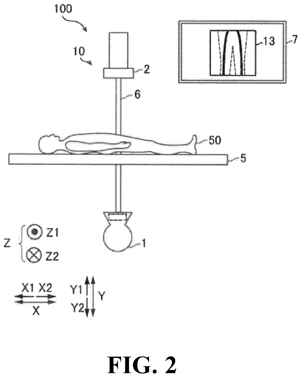
[0029]Referring to FIG. 1 to FIG. 10, the configuration of the X-ray fluoroscopic imaging apparatus 100 according to this embodiment will be described. Note that the X-ray fluoroscopic imaging apparatus 100 is an example of the “radiation fluoroscopic imaging apparatus” recited in claims.

(Configuration of X-Ray Fluoroscopic Imaging Apparatus)

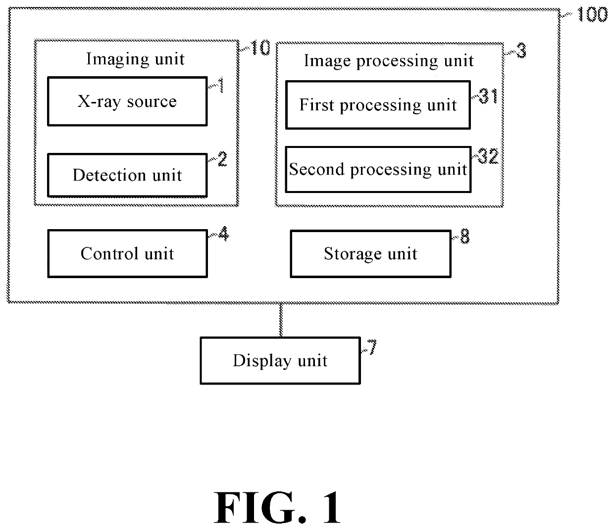
[0030]As shown in FIG. 1, the X-ray fluoroscopic imaging apparatus 100 of this embodiment is provided with an imaging unit 10 including an X-ray source 1 and a detection unit 2, an image processing unit 3, and a control unit 4.

[0031]As shown in FIG. 2, the X-ray fluoroscopic imaging apparatus 100 of this embodiment is configured to irradiate a subject 50 placed on a top board 5 with X-rays from an X-ray source 1.

[0032]The top board 5 is formed in a rectangular flat plate shape in a plan view. The subj...


PUM

PropertyMeasurementUnit
timeaaaaaaaaaa
shapeaaaaaaaaaa
voltageaaaaaaaaaa

Abstract

In this radiation fluoroscopic imaging apparatus, an image processing unit is configured to generate a first image by performing first image processing that clarifies a blood vessel as an imaging target when capturing a plurality of contrast agent images and generate a plurality of second images used to generate a differential long image by performing second image processing.

Description

CROSS-REFERENCE TO RELATED APPLICATIONS[0001]The related application number JP2019-071327, entitled “radiation fluoroscopic imaging apparatus”, filed on Apr. 3, 2019, and invented by David Thibault Pelletier, Naoki Hasegawa, Keiichi Tannno, and Takaya Yamamoto upon which this patent application is based is hereby incorporated by reference.BACKGROUND OF THE INVENTIONField of the Invention[0002]The present invention relates to a radiation fluoroscopic imaging apparatus and, more particularly, to a radiation fluoroscopic imaging apparatus for generating a differential long image.Description of the Background Art[0003]Conventionally, an X-ray diagnostic apparatus configured to generate a differential long image by irradiating a subject with X-rays has been known. Such a radiation fluoroscopic imaging apparatus is disclosed in, for example, Japanese Unexamined Patent Application Publication No. 2010-162278.[0004]In the X-ray image diagnostic apparatus described in the above-mentioned Jap...

Claims


Application Information

Patent Timeline
Patent Type & Authority Patents(United States)
IPC IPC(8): A61B6/00G06T7/00G06T5/00G01N23/04
CPCA61B6/5241A61B6/463A61B6/481A61B6/487A61B6/504A61B6/54G01N23/043G06T5/003G06T7/0012G01N2223/401G06T2207/10121G06T2207/30101A61B6/52G06T7/0016G06T2207/10016G06T2207/20224
Inventor THIBAULT PELLETIER, DAVIDHASEGAWA, NAOKITANNO, KEIICHIYAMAMOTO, TAKAYA
Owner SHIMADZU CORP
Features
  • R&D
  • Intellectual Property
  • Life Sciences
  • Materials
  • Tech Scout
  • Unparalleled Data Quality
  • Higher Quality Content
  • 60% Fewer Hallucinations
Learn More