Biopsy sampler

a sampler and biopsy technology, applied in medical science, surgery, vaccination/ovulation diagnostics, etc., can solve the problems of unusable tissue samples for histology and dangerous blood loss levels

Inactive Publication Date: 2002-04-25
BOSTON SCI SCIMED INC
View PDF0 Cites 256 Cited by
  • Summary
  • Abstract
  • Description
  • Claims
  • Application Information

AI Technical Summary

Problems solved by technology

Extensive bleeding can occur as a result of cold resecting, which can lead to dangerous blood loss levels.
The tissue sample, however, may be inadvertently cauterized when the resecting site is cauterized rendering the tissue sample unusable fo...

Method used


Image

  • Biopsy sampler
  • Biopsy sampler
  • Biopsy sampler

Examples


Embodiment Construction

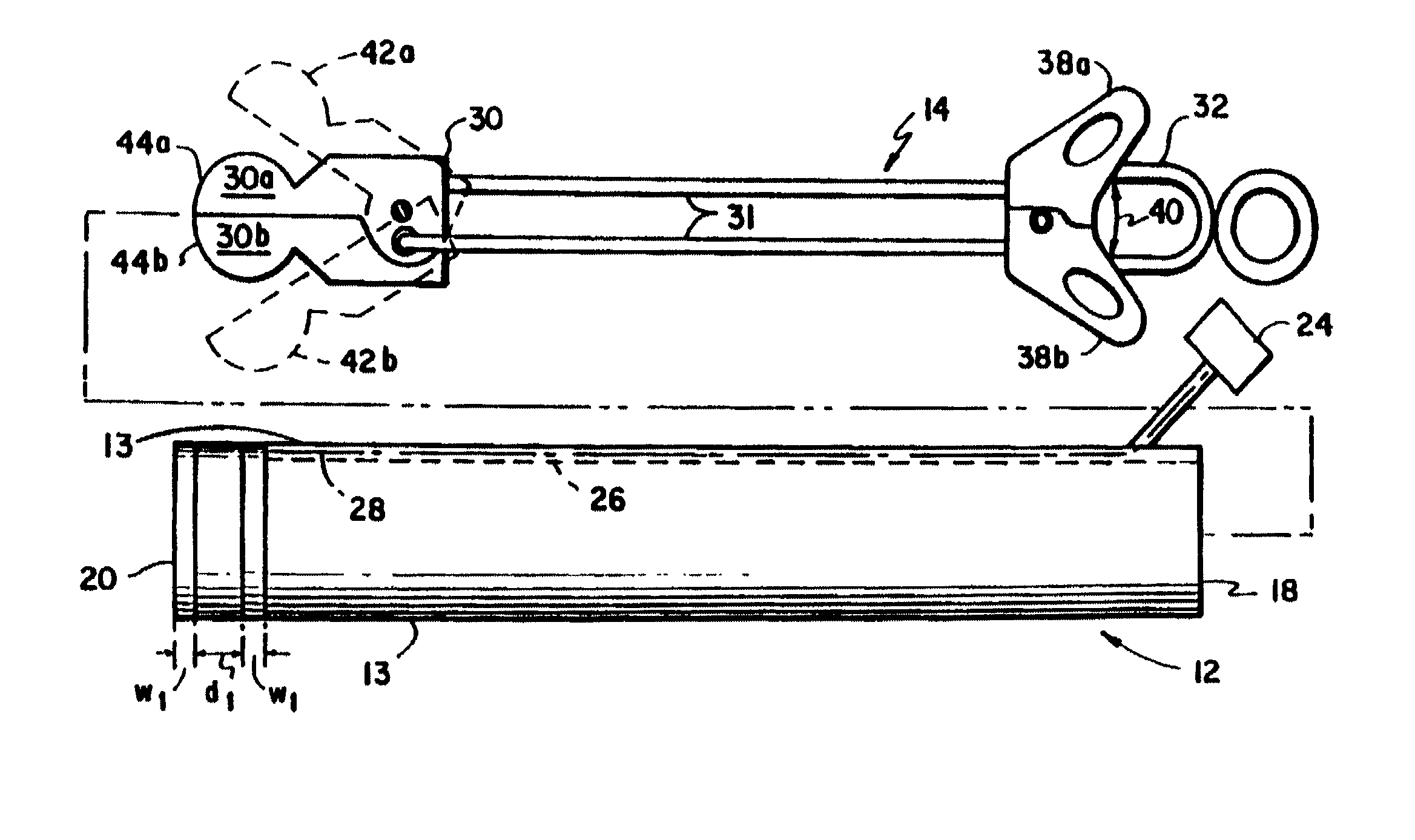
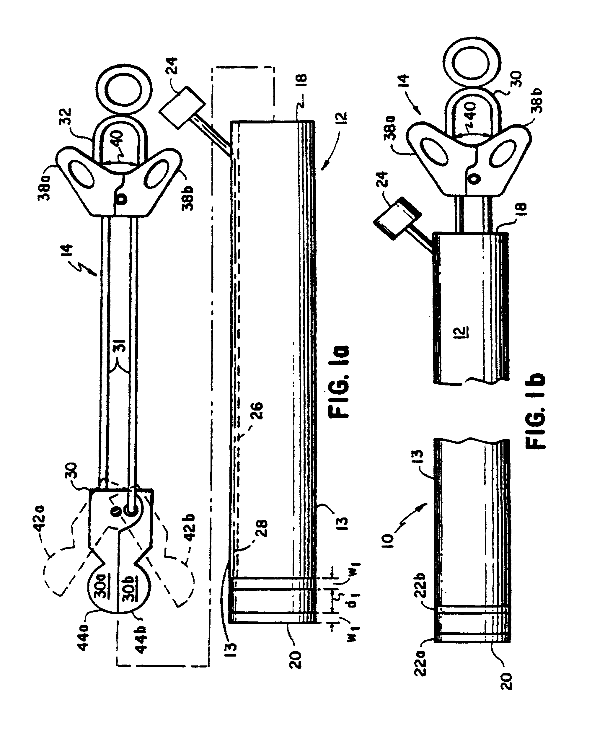
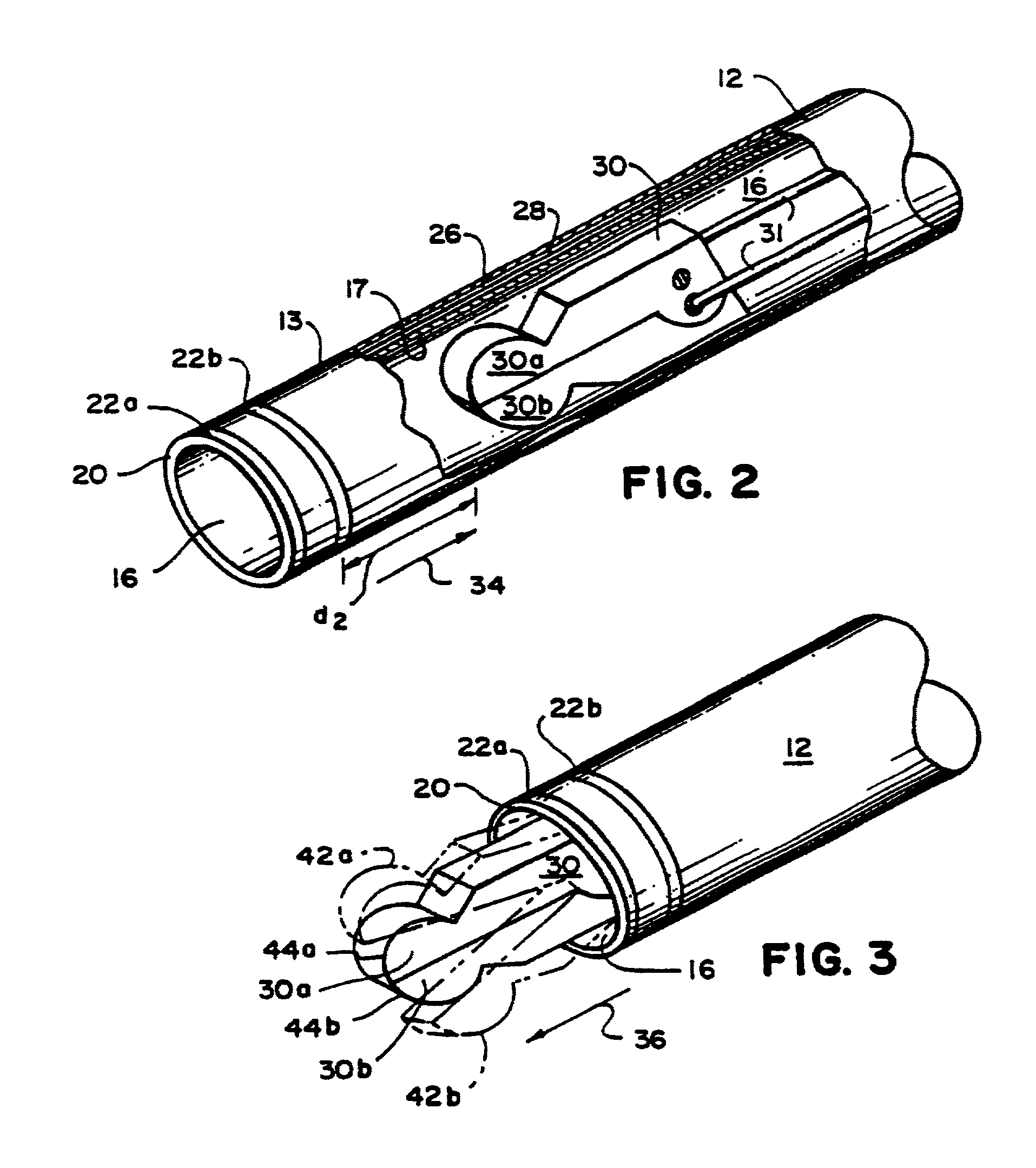
[0039] Referring to FIGS. 1a, 1b, 2, and 3, a biopsy assembly 10 includes a cauterizing sheath 12 and a resecting device 14. Sheath 12 includes a working lumen 16 extending from a proximal end 18 to a distal end 20 defined by an inner lumen wall 17 and sized to receive resecting device 14. The sheath also includes a pair of bipolar electrodes 22a, 22b mounted on an outer sheath surface 13 near distal end 20. Sheath 12 also includes an electrical connector 24, for connection to a radio frequency (RF) generator (not shown), and a lumen 26, extending between outer surface 13 and inner surface 17 of sheath 12 from electrical connector 24 to electrodes 22a, 22b. Two wires 28 are disposed within lumen 26, and each wire 28 electrically connects one of the electrodes 22a and 22b to electrical connector 24. Wires 28 are each coated with a layer of electrical insulation to prevent electrical contact between the wires.

[0040] Resecting device 14 is slidably positioned within working lumen 16 an...


PUM


Abstract

The invention features an assembly for taking a biopsy sample from a site within the body of a patient. The assembly includes a resecting device having a cutter near its distal end for resecting and containing a tissue sample and a sheath exterior to the resecting device and sized to be present within the body with the resecting device. The sheath includes an electrode element electrically isolated from the resecting device and disposed on the sheath's outer surface for cauterizing tissue. The electrode element may reside on the outer sheath, the distal end or both the outer sheath and the distal end of the assembly. The resecting device and the sheath cooperate to permit sequential resecting of a tissue sample from a resecting site and cauterizing of the site with the cutter sufficiently spaced from the electrode element to avoid heat damage to the tissue sample.

Description

[0001] This application is a continuation-in-part of Ser. No. 09 / 361,532 which was filed on Jul. 27, 1999 entitled Biopsy Sampler and is assigned to the same assignee as the present invention.[0002] 1. Field of the Invention[0003] This invention relates to biopsy sampling.[0004] 2. Description of Related Art[0005] There are many biopsy procedures in which tissue samples are taken for later histology. In "cold" biopsy procedures, surgical forceps are inserted within, for example, an endoscope, and are used to resect (i.e., cut) a tissue sample from, for instance, the biliary tree or colon. The forceps, and, hence, the tissue sample, are then removed from the patient through the endoscope.[0006] Extensive bleeding can occur as a result of cold resecting, which can lead to dangerous blood loss levels. Hence, after the tissue sample is removed, another device can be inserted into the patient through the endoscope to stop the bleeding. These devices include tamponade devices which apply ...

Claims


Application Information

Patent Timeline
IPC IPC(8): A61B10/00A61B10/06A61B17/32A61B17/34A61B18/14
CPCA61B10/02A61B10/06A61B17/32056A61B17/3478A61B2018/126A61B18/1492A61B2018/0016A61B2018/00595A61B18/1445
Inventor ROBERTS, TROY W.DIAMOND, BRUCE H.SHAW, WILLIAM J.
Owner BOSTON SCI SCIMED INC
Why Eureka
Try Eureka