Differentiation of human embryonic stem cells in avian embryos

a technology of embryonic stem cells and embryos, which is applied in the field of differentiation of human embryonic stem cells in avian embryos, can solve the problems of human cell types not yet produced, limitations of in vitro approaches,

Inactive Publication Date: 2005-04-21
YISSUM RES DEV CO OF THE HEBREWUNIVERSITY OF JERUSALEM LTD +1
View PDF0 Cites 5 Cited by
  • Summary
  • Abstract
  • Description
  • Claims
  • Application Information

AI Technical Summary

Benefits of technology

The present invention provides a way to test if something is harmful for humans by using special types of stem cells taken from early-stage fetuses. These cells are exposed to different substances and their response tells us about how they would be affected in real life. This can help identify potential hazards before products containing them make it onto store shelves.

Problems solved by technology

The patent text describes a method for obtaining normal differentiated human cells that can be used for therapeutic transplantation and pharmacological testing. The technical problem addressed is the limitations of current methods for in vitro differentiation of human embryonic stem cells, which can produce some but not all types of cells needed for patient needs or drug testing. The method involves inducing differentiation of human embryonic stem cells in a chick embryo system, which provides the correct combination of growth factors and extracellular matrix components needed for differentiation. The resulting cells can be recovered and used for further applications.

Method used


Image

  • Differentiation of human embryonic stem cells in avian embryos
  • Differentiation of human embryonic stem cells in avian embryos
  • Differentiation of human embryonic stem cells in avian embryos

Examples


example 1

Human ES Cells Transplanted into Somitic Mesoderm Integrates into Chick Tissues

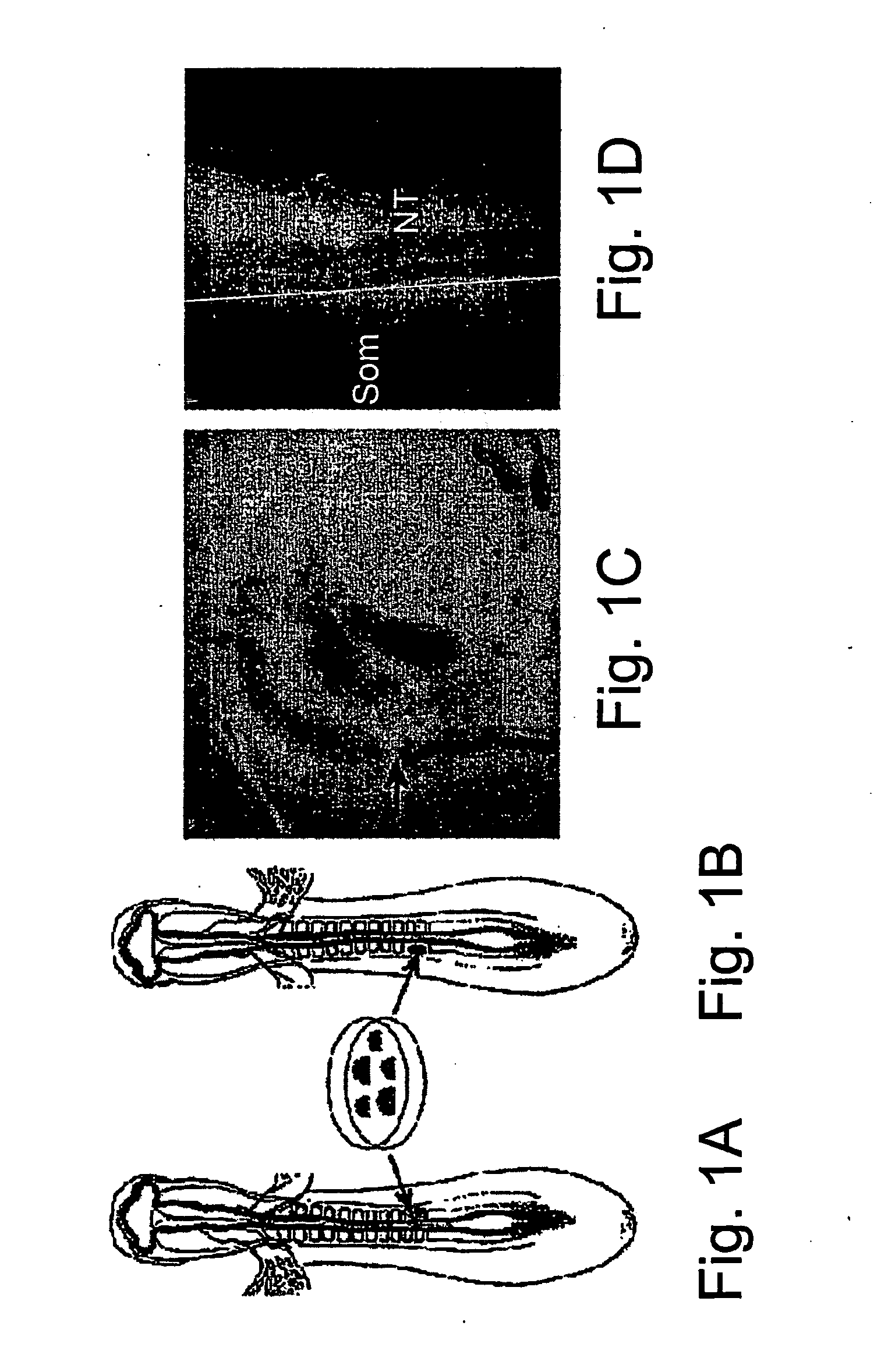
[0123] Colonies of human ES cells were micro-surgically grafted into the trunk region of 1.5 or 2 day-old (E1.5-E2) chick embryos (FIGS. 1A and 1B). One day after surgery the operation site was always visible. When GFP-expressing cells (Eiges et al. 2001) were implanted, they were clearly visible in the living embryo using fluorescence illumination (FIGS. 1C and 1D). The cells remained mostly as clumps, although individual cells could sometimes be observed migrating away from the site of implantation (not shown). The graft could be observed by fluorescence microscopy in some cases as long as four or five days post-surgery, after fixation and removal of overlying tissues (not shown).

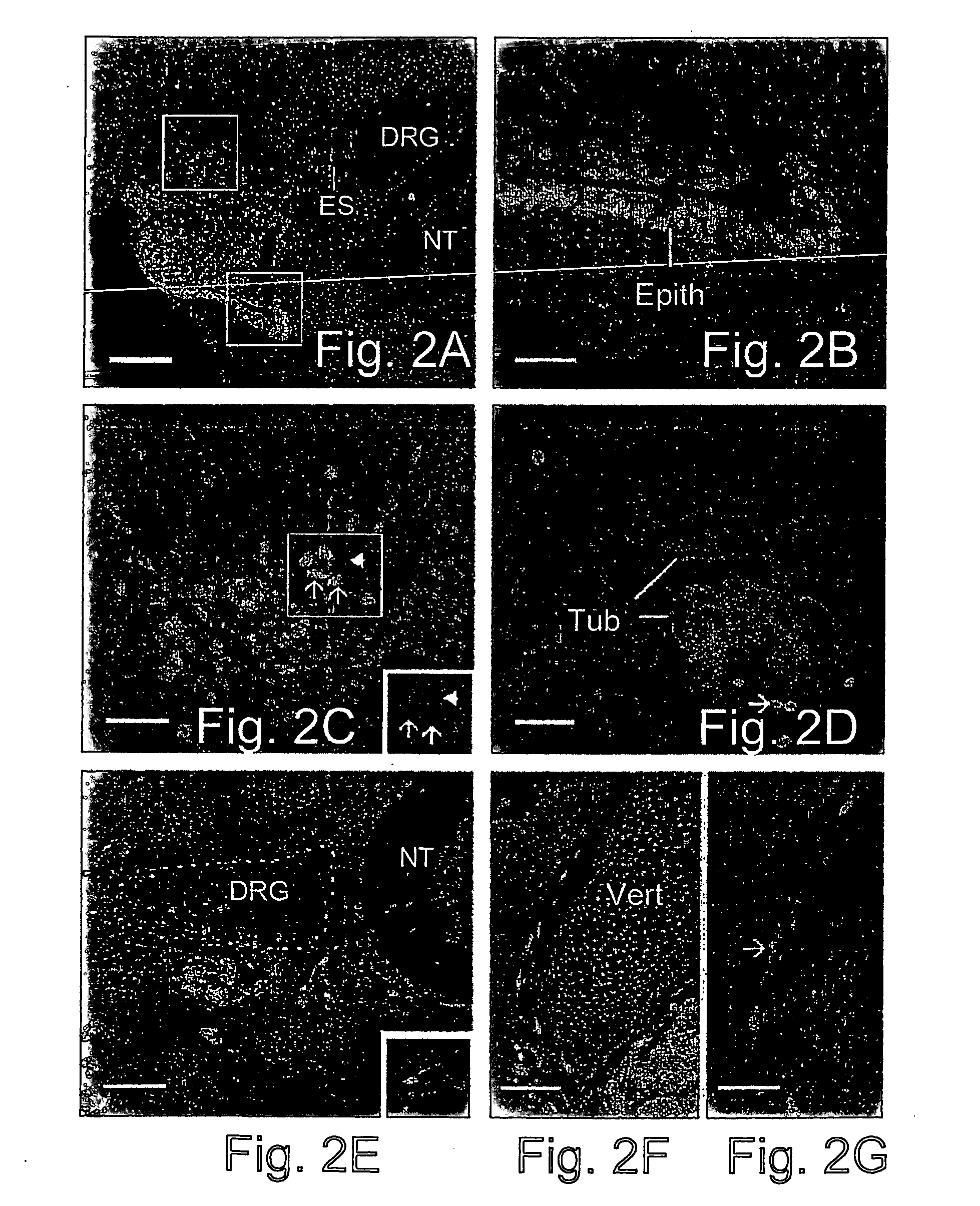
[0124] The somites give rise to multiple tissue types, including muscle, dermis and cartilage / bone. In addition, neural crest cells forming peripheral ganglia migrate through the somites after their epithelial / mesenchymal tr...

example 2

Neuronal Differentiation of Human ES Cells Replacing Somitic Mesoderm

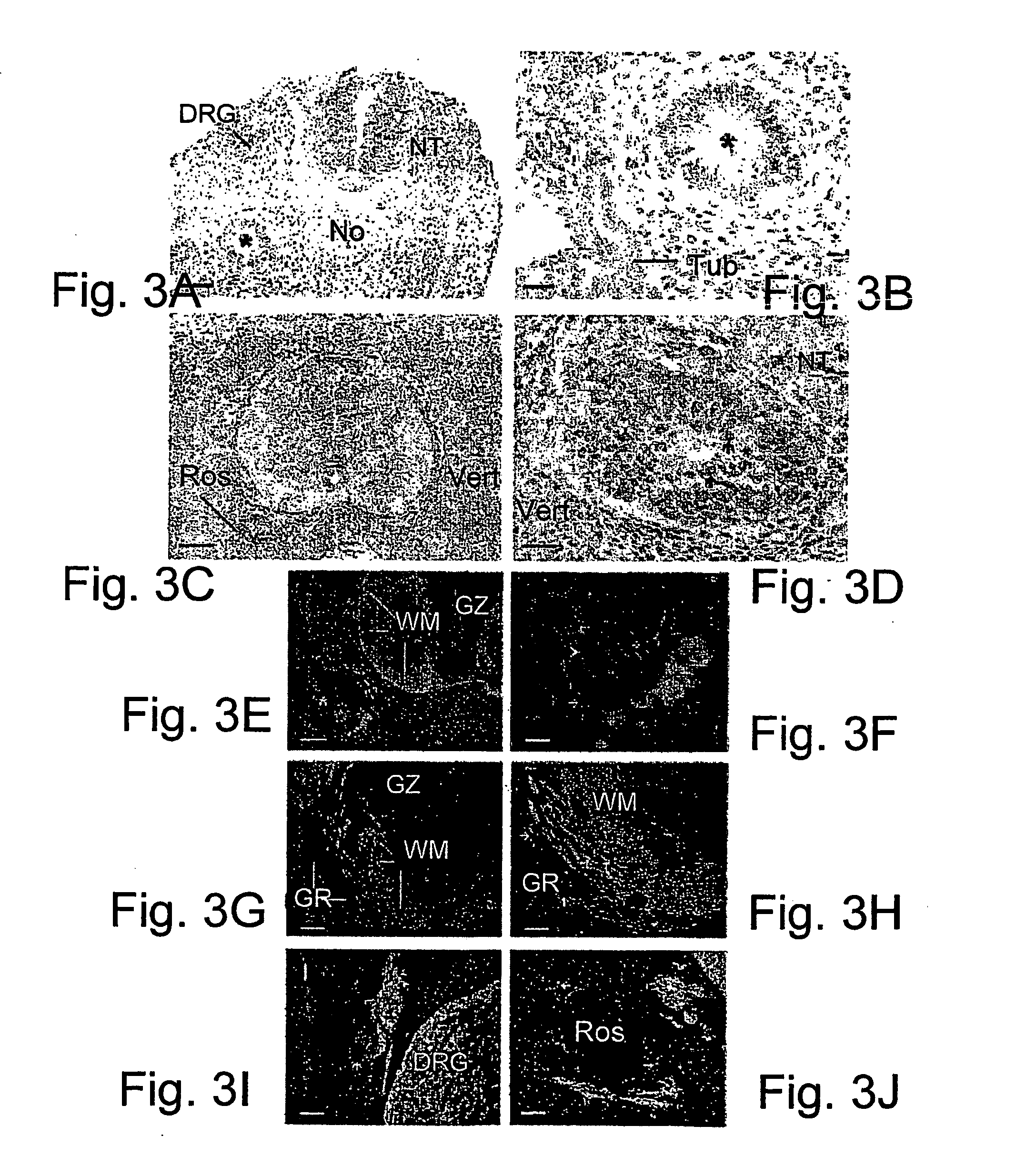
[0126] When colonies of human ES cells were implanted adjacent to the neural tube and notocord without intervening somitic mesoderm, epithelia reminiscent of neural rosettes were always (7 of 7 embryos analyzed) observed latero-ventral to the chick spinal cord (FIGS. 3A-3F). At embryonic days 6-7 these structures contained numerous mitotic figures that were localized primarily to their lumenal aspect (FIG. 3D). This arrangement of a stratified (or pseudo-stratified) epithelium with mitotic figures adlumenal and not basal, is characteristic of neural rosettes in human teratomas (Caccamo et al., 1989) and in the early vertebrate neural tube (see below). The neural rosette-like structures contained nuclei that were much larger than those of the host chick cells (FIGS. 3A-D).

[0127] Compared to human ES cells transplanted into damaged somites, this series of grafts contained many fewer individual cells that migrated ...


PUM

PropertyMeasurementUnit
Timeaaaaaaaaaa
Timeaaaaaaaaaa
Timeaaaaaaaaaa

Abstract

The invention relates to a method of preparing from human embryonic stem cells, differentiated cells suitable for transplantation, by introducing human embryonic stems cells into an avian host embryo. Also provided is a method of directing the differentiation of human embryonic stem cells by introducing them into a selected location in an avian host embryo, which dictates their differentiation pattern. The invention provides normal, transplantable differentiated human cells, e.g. progenitor and other cells, particularly neural cells. The invention also relates to therapeutic and diagnostic methods employing the differentiated cells of the invention.

Description


Claims


Application Information

Patent Timeline
Owner YISSUM RES DEV CO OF THE HEBREWUNIVERSITY OF JERUSALEM LTD
Why Eureka
Try Eureka