Methods and systems for the detection of malignant melanoma

Inactive Publication Date: 2005-10-13
DUKE UNIV
View PDF6 Cites 22 Cited by
  • Summary
  • Abstract
  • Description
  • Claims
  • Application Information

AI Technical Summary

Benefits of technology

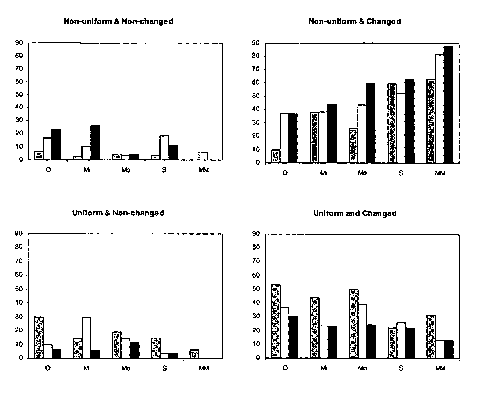
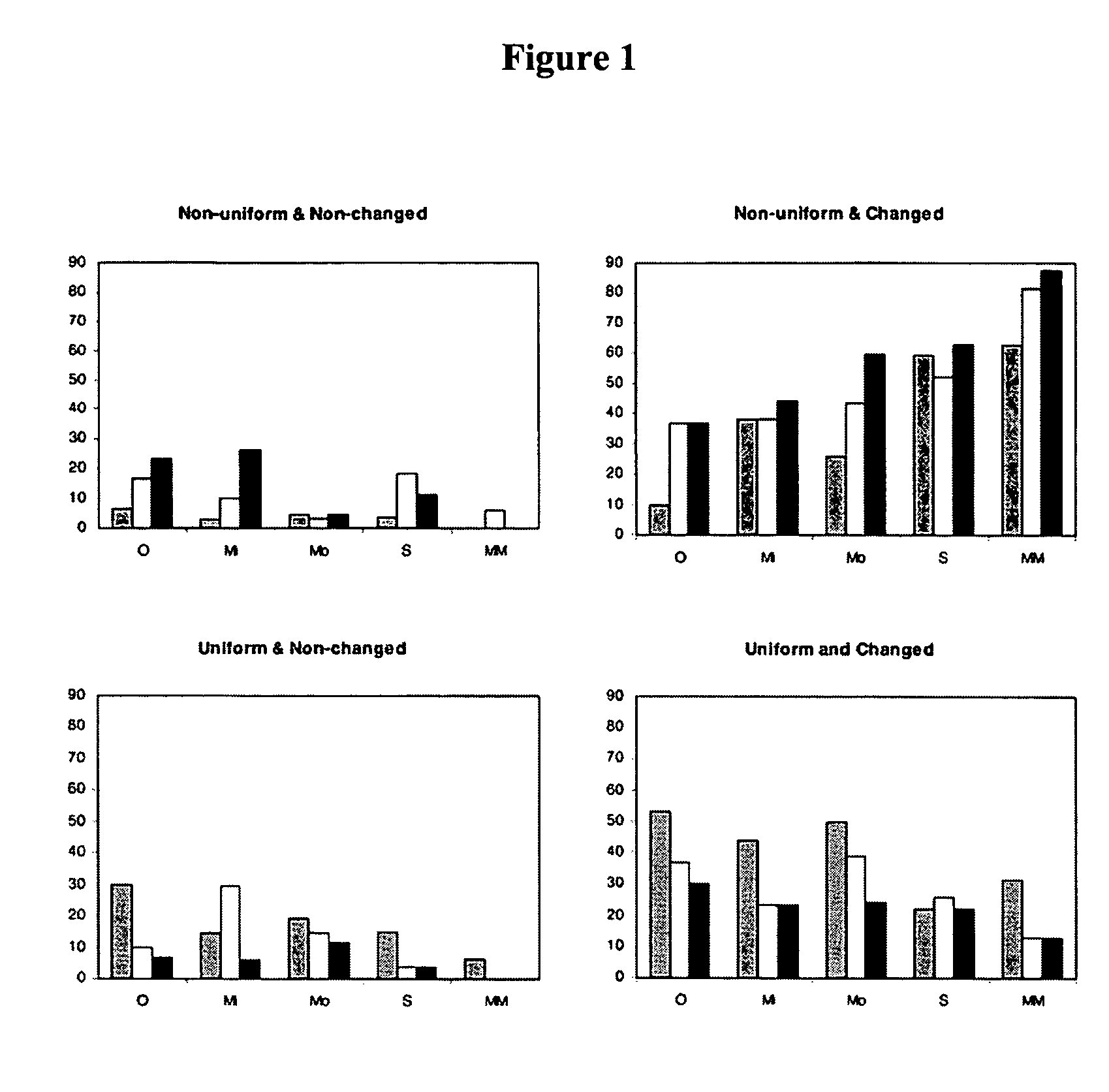
[0009] Accordingly, in some embodiments, the present invention provides a method, comprising the steps of: providing at least one initial body image (e.g., photograph or digital image) from a subject; and at least one follow up body image from the subject, wherein the follow up body image is taken at a later time than the initial body image; and comparing the initial body image and the follow up body image to identify moles or lesions that are both nonuniform and changed. In some embodiments, the body images are in a 2 or 3 dimensional format (e.g., obtained through cameras or scanning devices). In some embodiments, the method further comprises the step of providing a diagnosis of the presence or absence of melanoma in the subject. In other embodiments, the method further comprises the step of providing a level of the subject's risk of developing melanoma. In some embodiments, moles or lesions that are both nonuniform and changed are indicative of potential melanoma. In other embodiments, moles or lesions that are both nonuniform and changed are indicative of dysplastic nevus with severe cytologic atypia. The present invention is not limited to a particular interval of follow up images. However, in certain preferred embodiments, the at least one fo

Problems solved by technology

Studies show that distinguishing between benign pigmented lesions and early melanomas can be difficult, and

Method used


Image

  • Methods and systems for the detection of malignant melanoma
  • Methods and systems for the detection of malignant melanoma

Examples


example

Early Detection of Melanoma

[0059] This example describes methods for the early detection of melanoma utilized in some embodiments of the present invention.

A. Materials And Methods

Data Set

[0060] All lesions suggestive of melanoma are removed on a patient's initial visit. None of these lesions are included in this set. Patients perceived as being at high risk (numerous clinically atypical [dysplastic] nevi or prior melanoma) are then photographed (high-resolution digital 3072×2048, 33 views) (MoleMapCD, DigitalDerm Inc) and followed up. A 4-step process is then undertaken that is efficient and used on all follow up visits. First, lesions that concern the patient are identified. Second, total body skin examination is undertaken to identify lesions with concerning clinical features or lesions appearing to deviate from the patient's average type(s) of mole(s). Third, dermoscopy is used for worrisome lesions identified during the first 2 steps. Fourth, all lesions for which there i...


PUM


Abstract

The present invention relates to the detection of malignant melanoma. In particular, the present invention relates to systems and methods for the early detection of cancerous lesions.

Description

[0001] This application claims priority to provisional patent application Ser. No. 60 / 561,570, filed on Apr. 13, 2004, which is herein incorporated by reference in its entirety.FIELD OF THE INVENTION [0002] The present invention relates to the detection of malignant melanoma. In particular, the present invention relates to systems and methods for the early detection of cancerous lesions. BACKGROUND OF THE INVENTION [0003] Melanoma is a malignant tumor of melanocytes, cells that are derived from the neural crest. Although most melanomas arise in the skin, they may also arise from mucosal surfaces or at other sites to which neural crest cells migrate. Melanoma occurs predominantly in adults, and more than half of the cases arise in apparently normal areas of the skin. Early signs of malignant melanoma in a mole include darker or variable discoloration, itching, or an increase in size. Ulceration or bleeding are later signs. Melanoma can arise from any site on the skin surface. [0004] ...

Claims


Application Information

Patent Timeline
IPC IPC(8): A61B5/00A61B5/05A61B5/103A61B8/00A61B10/00G06F17/00G06T7/00
CPCA61B5/0059A61B5/415A61B5/418G06T2207/30024A61B5/445A61B5/7275G06T7/0012A61B5/444G16H50/30G16H30/40G16H50/20G06V20/698
Inventor GRICHNIK, JAMES M.
Owner DUKE UNIV
Why Eureka
Try Eureka