Methods of perfusion imaging

a perfusion imaging and magnetic resonance technology, applied in the field of magnetic resonance imaging, can solve problems such as the association of blood perfusion deficits

Inactive Publication Date: 2006-06-08
CYTOGENESIS INC +2
View PDF12 Cites 15 Cited by
  • Summary
  • Abstract
  • Description
  • Claims
  • Application Information

AI Technical Summary

Benefits of technology

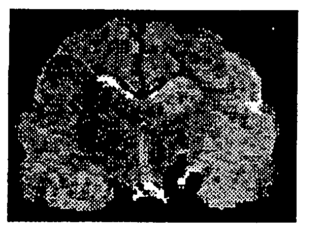
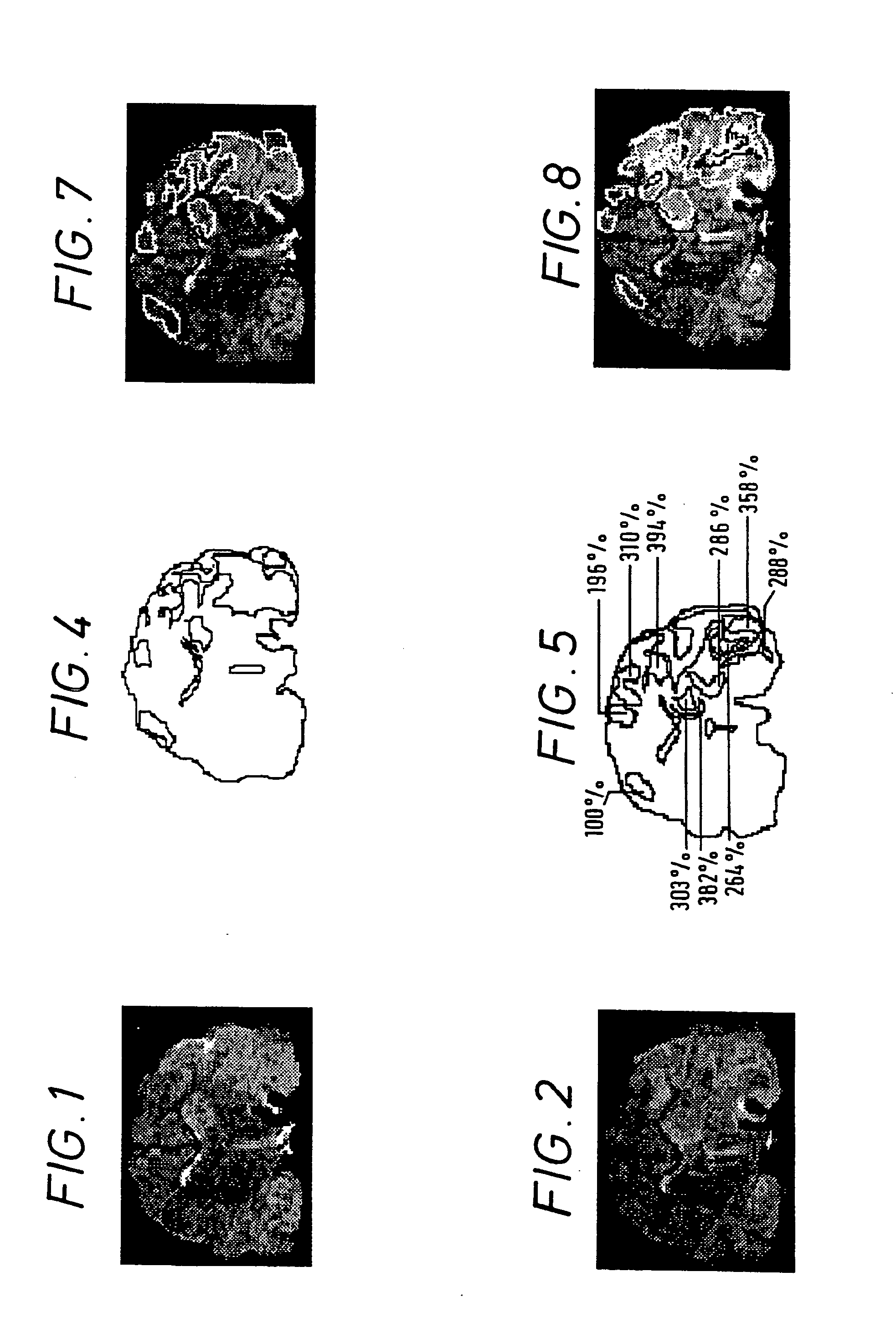
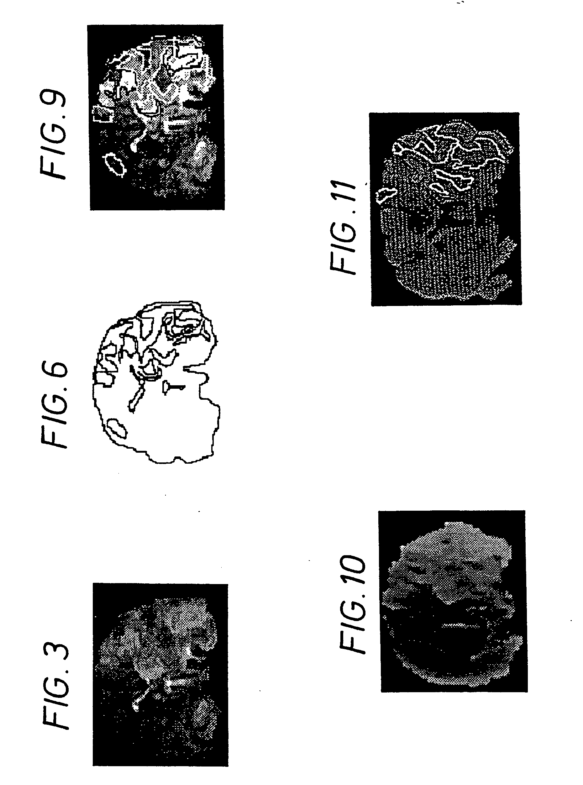
[0013] In one embodiment of the method of the invention, using a fast imaging procedure, the determination of the location and severity of ischaemia is effected by determining the time dependence of the MR signal intensity for the voxels in the seconds following administration of the contrast agent, and generating an image where voxel image intensity value is dependent on the time post-administration at which MR signal intensity for that voxel is lowest. Normal tissue reaches minimum MR signal intensity sooner than ischaemic tissue, and the resulting image thus enables the spatial extent and local severity of blood flow abnormality to be visualized. Alternatively, a similar image may be generated by making the voxel image intensity value dependent on the time taken before voxel MR signal intensity reattains a pre-selected control value, e.g., its pre-injection value or a percentage of that value (for example 80%).

Problems solved by technology

Blood perfusion deficits are associated with several serious and often life-threatening conditions.

Method used


Image

  • Methods of perfusion imaging
  • Methods of perfusion imaging
  • Methods of perfusion imaging

Examples


Embodiment Construction

[0044] The magnetic susceptibility, T2*-reducing effect, of MS contrast agents is to a large degree dependant on the magnitude of the magnetic moment of the magnetic species within the contrast agent—the higher the magnetic moment the stronger the effect. Indeed the effect is approximately proportional to the square of the magnetic moment making the effect of Dy(III) about 1.95 times larger than that of Gd(III). In general paramagnetic metal species having magnetic moments of ≧4 BM will be preferred. The contrast agents particularly preferred for use in the method of the present invention are those containing paramagnetic lanthanide ions, especially high spin lanthanides such as ions of Dy, Gd, Eu, Yb and Ho, in particular Dy(III).

[0045] In order that they may be administered at effective but non-toxic doses, such paramagnetic metals will generally be administered in the form of ionic or much more preferably non-ionic, complexes, especially chelate complexes optionally bound to lar...


PUM


Abstract

The invention provides a method of imaging a blood vessel in a body using magnetic resonance (MR) imaging. The method includes the step of acquiring images of a region of the body in which the blood vessel is located. It also includes the step of administering an MR contrast agent into the vascular system of the body at a dose and a time selected so that the MR contrast agent passing through the blood vessel is present therein during acquisition of the images thereof for at least part of the acquiring step.

Description

CROSS-REFERENCE TO RELATED APPLICATIONS [0001] This is a continuation of U.S. application Ser. No. 09 / 189,043 (filed 9 Nov. 1998), which is a continuation of U.S. application Ser. No. 08 / 604,778 (filed 23 Feb. 1996, now U.S. Pat. No. 5,833,947), which is continuation of U.S. application Ser. No. 08 / 306,221 (filed 14 Sep. 1994, now U.S. Pat. No. 5,494,655), which is a continuation of U.S. application Ser. No. 07 / 946,373 (filed 30 Oct. 1992, abandoned). U.S. application Ser. No. 07 / 946,373 was filed pursuant to 35 U.S.C. §371 as a national stage application derived from PCT International Application No. PCT / EP91 / 00443 (filed 6 Mar. 1991), and is a continuation of U.S. application Ser. No. 07 / 490,859 (filed 9 Mar. 1990, now U.S. Pat. No. 5,190,744).FIELD OF THE INVENTION [0002] This invention relates to improvements in and relating to magnetic resonance imaging, in particular imaging of phenomena associated with blood flow variations and abnormalities. BACKGROUND OF THE INVENTION [0003...

Claims


Application Information

Patent Timeline
Patent Type & Authority Applications(United States)
IPC IPC(8): A61B5/05
CPCA61B5/0263A61B5/0275G01R33/5601G01R33/563G01R33/56366
Inventor KUCHARCZYK, JOHNMOSELEY, MICHAEL E.
Owner CYTOGENESIS INC
Why Eureka
Try Eureka