Sheath with channel for endoscope

a technology of endoscope and sheath, which is applied in the field of sheaths for medical devices, can solve the problems of enlarge the cross-section of the sheath-covered endoscope being inserted into the patient, affecting the patient's health, and affecting the patient's overall health,

Inactive Publication Date: 2007-06-21
COGENTIX MEDICAL
View PDF19 Cites 155 Cited by
  • Summary
  • Abstract
  • Description
  • Claims
  • Application Information

AI Technical Summary

Benefits of technology

[0016] An aspect of some embodiments of the present invention relates to an endoscopic sheath adapted to allow insertion of a working tube into a channel defined by the sheath. The working tube defines an external protrusion and the sheath includes a respective groove for receiving the protrusion, running along its length. In an exemplary embodiment of the invention, the external protrusion comprises a dovetail and the groove comprises a partially closed notch which prevents annular escape of the protrusion from the groove. Alternatively, annular escape is prevented by the tight fitting of the working tube into the relatively flexible channel. The groove may run over the entire length of the sheath or may run over a portion (e.g., a proximal portion) of the sheath.
[0017] An aspect of some embodiments of the present invention relates to a method of inserting a working tube into a channel defined by an endoscopic sheath with a variable transverse extent, using a guide wire running along the channel. In some embodiments of the invention, the guide wire is preinserted in the channel, for example at the time of manufacture of the sheath, so that there is no need to separately insert the guide wire into the patient. Alternatively, the guide wire is inserted into the channel after the sheath is inserted into the patient, so that the guide wire does not interfere in inserting the sheath into the patient.

Problems solved by technology

This, however, enlarges the cross-section of the sheath-covered endoscope being inserted into the patient.
Such a larger diameter may make the insertion of the endoscope more difficult or may prevent the insertion altogether.
The inflating of the channel is problematic, however, especially when the endoscope is inserted to a tight tissue location.
Furthermore, even if the inflating of the channel is possible, the forces involved in inflating the channel may distort the layout of the endoscope.

Method used


Image

  • Sheath with channel for endoscope
  • Sheath with channel for endoscope
  • Sheath with channel for endoscope

Examples


Embodiment Construction

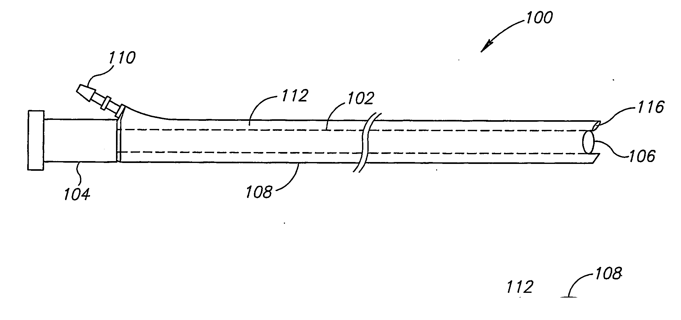
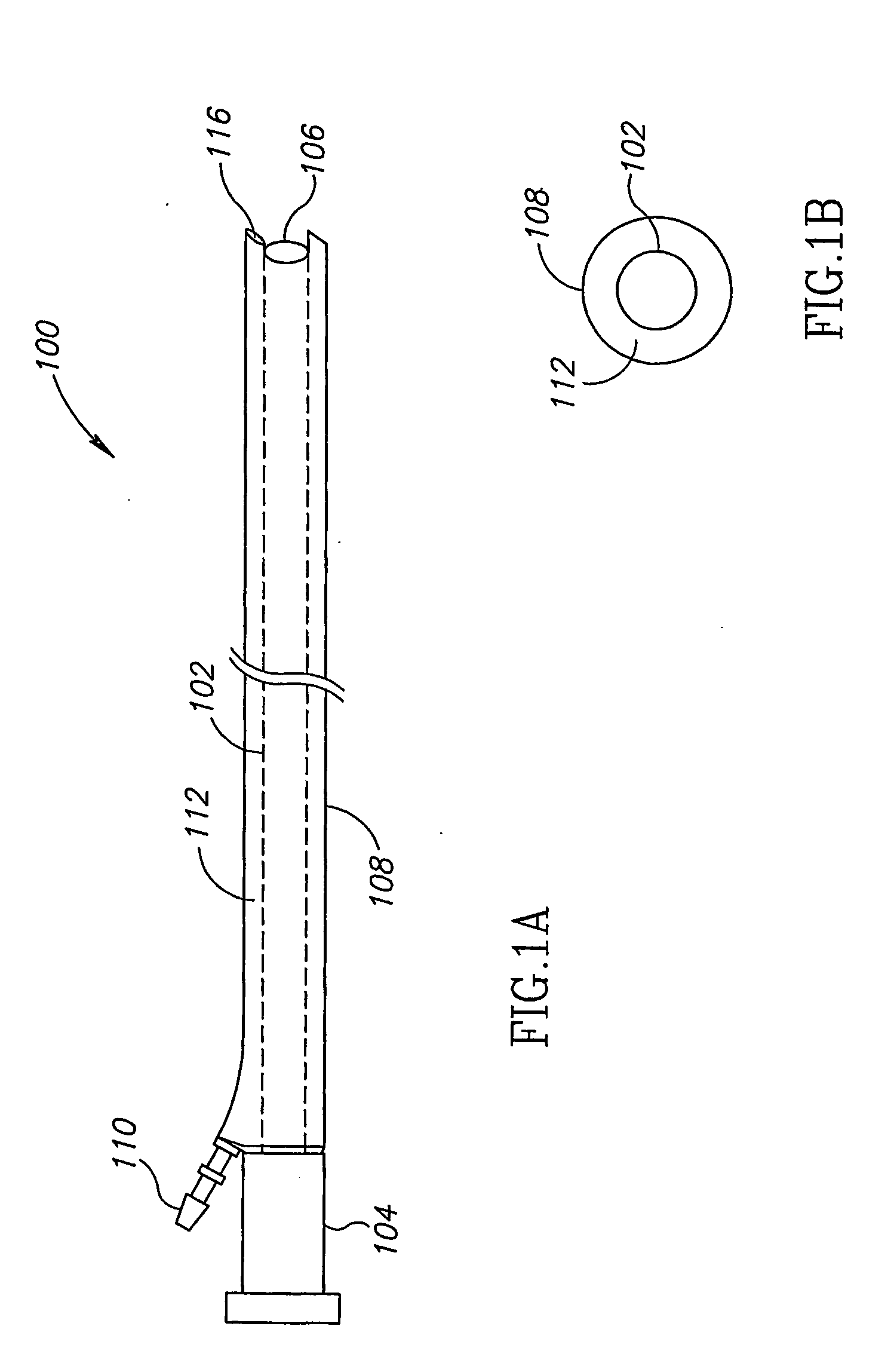
[0064]FIGS. 1A and 1B are a schematic side view and a cross sectional view of a sheath assembly 100, in accordance with an exemplary embodiment of the present invention. Assembly 100 optionally includes an internal sheath 102 adapted to receive an endoscope and isolate the endoscope from the environment. In some embodiments of the invention, a rigid pipe section 104 is located at a proximal end of internal sheath 102, to aid insertion of the endoscope into the sheath. A sealed window 106 at the distal end of internal sheath 102, optionally isolates the endoscope from the environment while allowing a camera or fiberoptic image bundle of the endoscope to provide images of the tissue external to sheath assembly 100.

[0065] An external sheath 108, having a larger circumference than internal sheath 102, optionally surrounds internal sheath 102. During insertion of an endoscope with sheath assembly 100 into a patient, external sheath 108 is optionally closely folded around internal sheath...


PUM


Abstract

A sheath assembly (100) for an invasive probe. The sheath assembly (100) includes an internal sheath (102) for covering a probe and at least one channel tube (112) external to the internal sheath (102), the channel tube (112) being foldable into a closed state in which the tube does not define a channel, or openable into an open state in which the tube defines a channel that extends along a portion of the sheath assembly (100).

Description

RELATED APPLICATIONS [0001] This application claims the benefit under 119(e) of U.S. provisional application No. 60 / 491,971, filed Aug. 4, 2003, the disclosure of which is incorporated herein by reference.FIELD OF THE INVENTION [0002] The present invention relates to sheaths for medical apparatus. BACKGROUND OF THE INVENTION [0003] Endoscopes are used to view internal tissue of humans, and for many other tasks. As sterilization of endoscopes is relatively difficult, disposable sheaths which cover an endoscope are used to isolate the endoscope from the patient tissue, so as to avoid time-consuming cleaning and disinfection processes. In some cases it is desired to have one or more channels run along the endoscope. These channels may be used, for example, to pass tools and fluids to the tip of the endoscope. As the sheath should completely isolate the endoscope from the human tissue, such channels are generally attached to the sheath, so that they are on an outer side of the endoscope...

Claims


Application Information

Patent Timeline
Patent Type & Authority Applications(United States)
IPC IPC(8): A61B1/00A61F
CPCA61B1/00135A61B1/0014A61B1/00142A61B2019/082A61B1/015A61B1/018A61B1/012A61B46/10A61B46/13
Inventor MARTONE, STEPHENHADANI, RON
Owner COGENTIX MEDICAL
Why Eureka
Try Eureka