Method for examining kidney diseases
a kidney disease and kidney prognosis technology, applied in the field can solve the problems of not being able to establish a decisive method for accurate diagnosis of kidney disease prognosis, and aiming at the existence of such a method in the tissues, and achieve the effect of improving the prognosis
- Summary
- Abstract
- Description
- Claims
- Application Information
AI Technical Summary
Benefits of technology
Problems solved by technology
Method used
Image

Examples
example 1
Preparation (I) of Antibody Binding to FABP in Human Kidney Tissue (Preparation of Anti-mouse FABP Antibody):
(1) Anti-mouse L-FABP Polyclonal Antibody:
[0060] FABP existing at the human proximal tubule has been known to be mainly a liver-type FABP (L-FABP). Human L-FABP and mouse L-FABP have a high homology, and as an antibody binding to L-FABP in human kidney tissues, anti-mouse L-FABP antibody may be used.
[0061] Thus, an anti-mouse L-FABP polyclonal antibody was prepared. The antigen, mouse L-FABP, was prepared according to the method disclosed in the literature (Takahashi et al., Eur. J. Biochem., vol. 136, p. 589-601, 1983), as follows. That is, to the excised liver from a mouse killed by bleeding was added a four-time volume of 30 mM Tris-HCl buffer (pH 8), and the mixture was treated by a polythoron-type homogenizer. The resultant was centrifuged at 8000 rpm for 15 minutes, and the supernatant thus obtained was further ultra-centrifuged at 100,000×g for 90 minutes to give ...
example 2
Preparation (II) of Antibody Binding to FABP in Human Kidney Tissues (Preparation of Anti-human L-FABP Antibody):
(1) Purification of Recombinant Human L-FABP:
[0065] cDNA of human L-FABP was obtained by PCR (polymerase chain reaction) from the .cDNA library derived from human liver (manufactured by CLONTECH Laboratories Inc., Cat # HL1115b Lot # 5621). An oligonucleotide of 23 to 27 mers synthesized by a DNA synthesizer was used as a primer. The necleotide sequence of the primer was designed based on the gene sequence of human L-FABP disclosed in the literature (Lowe et al., J. Biol. Chem., vol. 260, p. 3413-3417, 1985) and Gene Data Base (GENBANK Accession No. M10617), with adding a restriction enzyme recognition site for inserting an expression vector at the end of the primer. The obtained DNA fragment (about 420 base pairs) has a BamHI recognition site before the initiation codon, and the BamHI recognition site after the termination codon, and encodes the desired full-length h...
example 3
Localization of FABP in Human Kidney Tissues (Normal Kidney Tissues):
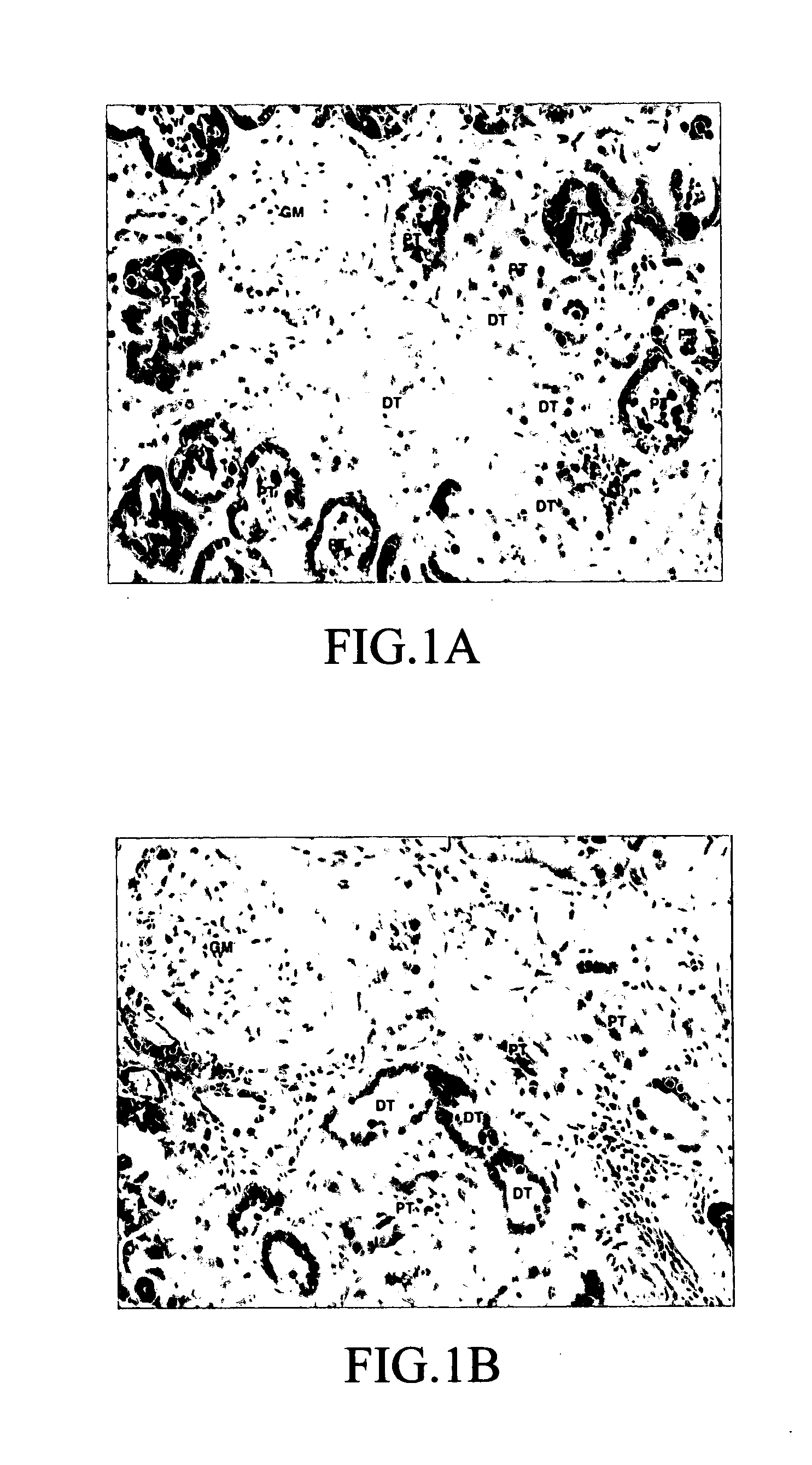
[0071] Normal human kidney tissues were subjected to immunohisto staining of FABP. The human kidney tissues were normal portions of the kidney excised from the patient with renal cancer. A primary antibody for L-FABP staining was the anti-mouse L-FABP polyclonal antibody (IgG) prepared in the same manner as in Example 1-(1). A primary antibody for H-FABP staining was the anti-mouse H-FABP polyclonal antibody (IgG) prepared in the same manner as in Example 1-(2). The immunostaining was carried out using Vectastain ABC kit (manufactured by Vector Laboratory, Inc.), and a secondary antibody was a biotinylated anti-rabbit IgG, and an enzyme was a biotinylated horseradish peroxidase, and a coloring substrate was DAB (3,3′-diaminobensidine tetrahydrochloride).
[0072] The results are shown in FIG. 1. When anti-L-FABP antibody was used, the proximal tubule was stained. On the other hand, when anti-H-FABP antibody was use...
PUM
 Abstract
Description
Claims
Application Information
 - R&D Engineer
- R&D Manager
- IP Professional
- Industry Leading Data Capabilities
- Powerful AI technology
- Patent DNA Extraction
Browse by: Latest US Patents, China's latest patents, Technical Efficacy Thesaurus, Application Domain, Technology Topic.
© 2024 PatSnap. All rights reserved.Legal|Privacy policy|Modern Slavery Act Transparency Statement|Sitemap