Integrated Multimodality Intravascular Imaging System that Combines Optical Coherence Tomography, Ultrasound Imaging, and Acoustic Radiation Force Optical Coherence Elastography

a multi-modality, intravascular imaging technology, applied in the field of multi-modality intravascular imaging systems, multi-modality endoscopic imaging systems, optical coherence elastography, acoustic radiation force systems, can solve the problems of increasing survival rate, reducing treatment, and current diagnostic techniques, including digital rectal exams and blood prostate-specific antigen (psa) measurement, can achieve improved prognosis, reduce costs for hospitals and patients, and improve sensitivity in displacement measuremen

Active Publication Date: 2015-12-10
RGT UNIV OF CALIFORNIA
View PDF4 Cites 34 Cited by
  • Summary
  • Abstract
  • Description
  • Claims
  • Application Information

AI Technical Summary

Benefits of technology

[0018]This multi-modal imaging strategy in a single system permits the use of a single disposable guide wire and catheter, thereby reducing costs to hospitals and patients, and improving prognosis by early detection.
[0019]The ARF-OCE combines the high-speed excitation of ARF with sub-micrometer / nanometer detection sensitivity of phase resolved OCT. This elastography technique allows high-speed and high-resolution point-by-point mapping of local strains in atherosclerotic coronary tissue, which provide critical information to assess the vulnerability of plaques.
[0021]A spectral and resonant PR-ARF-OCE technique, where a chirped ultrasound signal is used to generate a frequency sweep of ARF to measure the frequency dependence of OCE signal, can be used to measure quantitatively the mechanical properties of tissue and different tissues with both spectral and spatial resolution. This method takes advantage of the fact that materials response is enhanced at their mechanical resonant frequencies, which can be used effectively distinguish samples with varying stiffness.
[0025]The integrated intraluminal imaging system further includes a single disposable catheter into which the intraluminal probe is disposed, whereby costs are reduced and hence prognosis improved by early detection.
[0032]The method further includes the step of providing a single disposable catheter into which the intraluminal probe is disposed, whereby costs are reduced and hence prognosis improved by early detection.
[0035]The step of providing an acoustic generator generating an acoustic radiation force (ARF) signal coupled to the ultrasound transducer comprises generating a chirped amplitude modulated ultrasound beam to frequency sweep the acoustic radiation force delivered by the ultrasound transducer, where processing the returned optical signal, the returned ultrasound signal and the returned ARF optical signal comprising measuring frequency dependence of the returned ARF optical signal, and further comprising processing the measured frequency dependence of the returned ARF optical signal to quantitatively measure the mechanical properties of the identified tissues with both spectral and spatial resolution using enhanced materials response at mechanically resonant frequencies to distinguish tissues with varying stiffness.

Problems solved by technology

Early diagnosis of cancer increases the survival rate and reduces treatment.
Current diagnostic techniques, including digital rectal exam (DRE) and the measurement of blood prostate-specific antigen (PSA) levels, are insufficient for guiding treatment.
The chief prostate cancer imaging technique is ultrasound, but grayscale ultrasound imaging is only 50-60% accurate and transrectal ultrasound (TRUS) is even less accurate.
Ultrasound also has difficulty differentiating cancer from other diseases, such as benign prostatic hyperplasia (BPH) and prostatitis.
Despite advances in ultrasound techniques such as color and power Doppler, and the introduction of ultrasound contrast agents, TRUS is still limited in accuracy and therefore only used to guide biopsy.
Angioscopy allows the plaque to be visualized with high sensitivity, but the morphologic characterization of the plaque is unreliable because of the lack of estimation of cap thickness or lipid content.
Even though MRI is used to study the progression and regression of plaque over time, its insufficient resolution cannot render accurate measurements.
IVUS has limited resolution and sensitivity to assess the thickness of the thin fibrous cap and for plaque classifications.
OCT has a limited imaging depth and cannot quantify the full thickness of the plaque.
The limitation of NIRS, Raman and fluorescence imaging is that they lack the capability of tomographic imaging.
IVUS lacks the required high resolution to quantify the thickness of the fibrous cap.
However, this system lacks the capability to resolve tissue composition in plaque lesions.
Due to the small dimensions involved, time varying blood flow, and presence of multiple plaques of different sizes, geometries, and compositions, arterial elastography is more challenging than other elastographies such as breast tissue elastography.
The former applies compression to the subject statically / quasi-statically and measures the relaxation dynamics, which has limited imaging speed for in vivo imaging.
Phase-resolved dynamic OCE had been investigated previously for shear wave propagation detection, but this method requires separation between excitation and detection points, which limits the speed and spatial resolution.
Adaptation of this method for in vivo real time 2D and 3D vascular elastography is not viable.

Method used


Image

  • Integrated Multimodality Intravascular Imaging System that Combines Optical Coherence Tomography, Ultrasound Imaging, and Acoustic Radiation Force Optical Coherence Elastography
  • Integrated Multimodality Intravascular Imaging System that Combines Optical Coherence Tomography, Ultrasound Imaging, and Acoustic Radiation Force Optical Coherence Elastography
  • Integrated Multimodality Intravascular Imaging System that Combines Optical Coherence Tomography, Ultrasound Imaging, and Acoustic Radiation Force Optical Coherence Elastography

Examples


Embodiment Construction

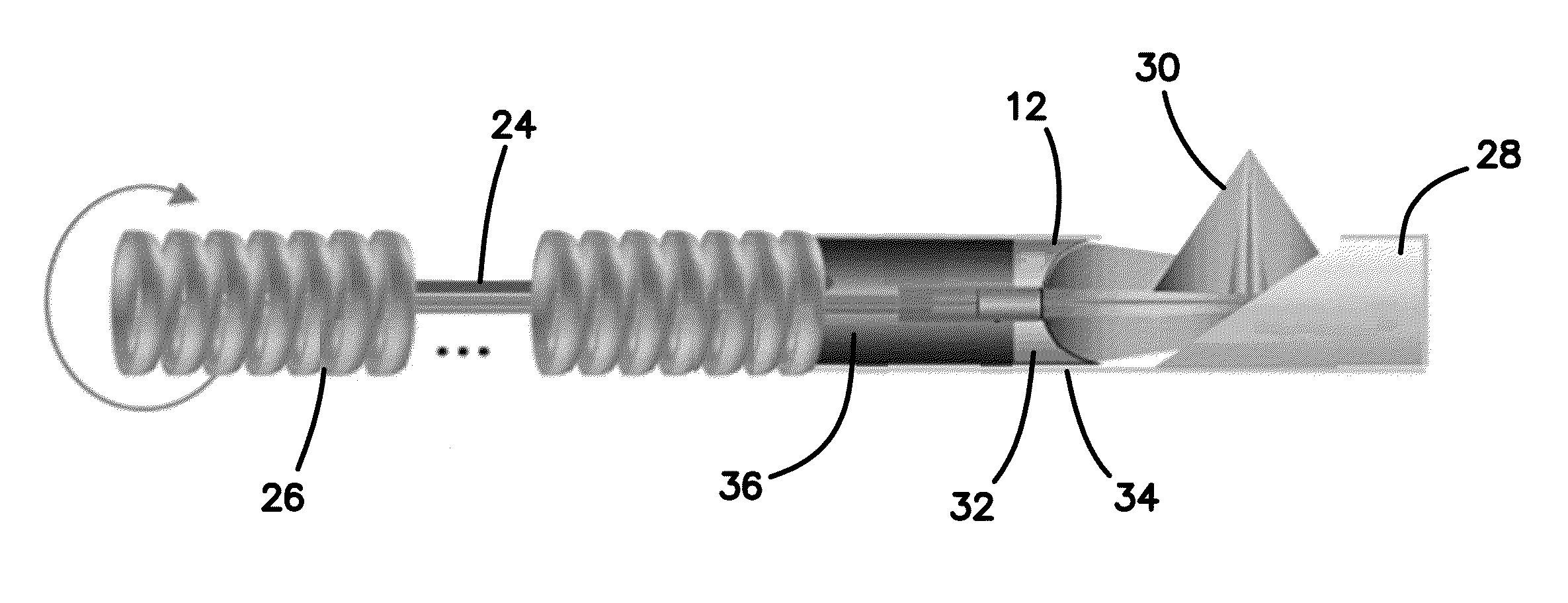
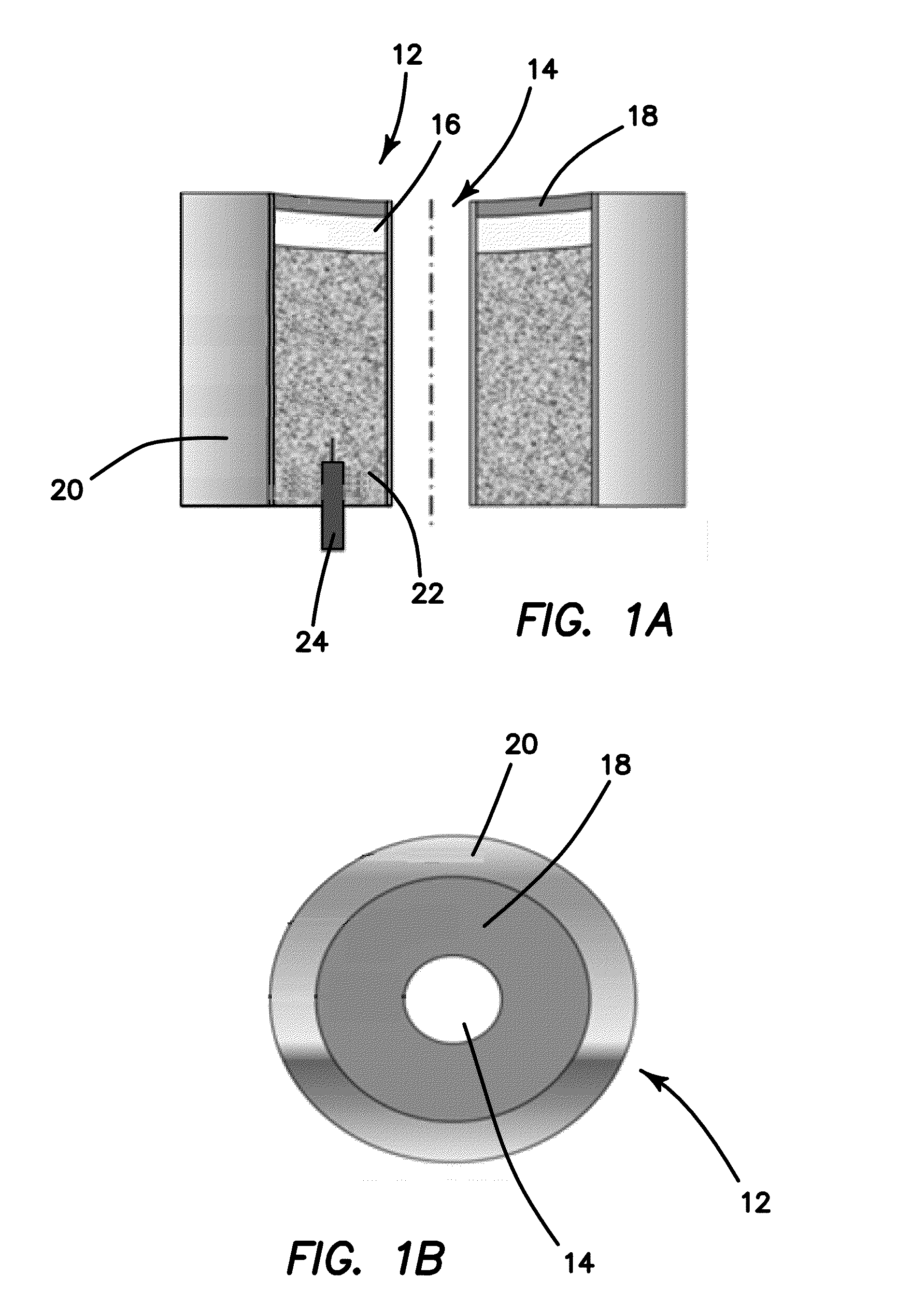
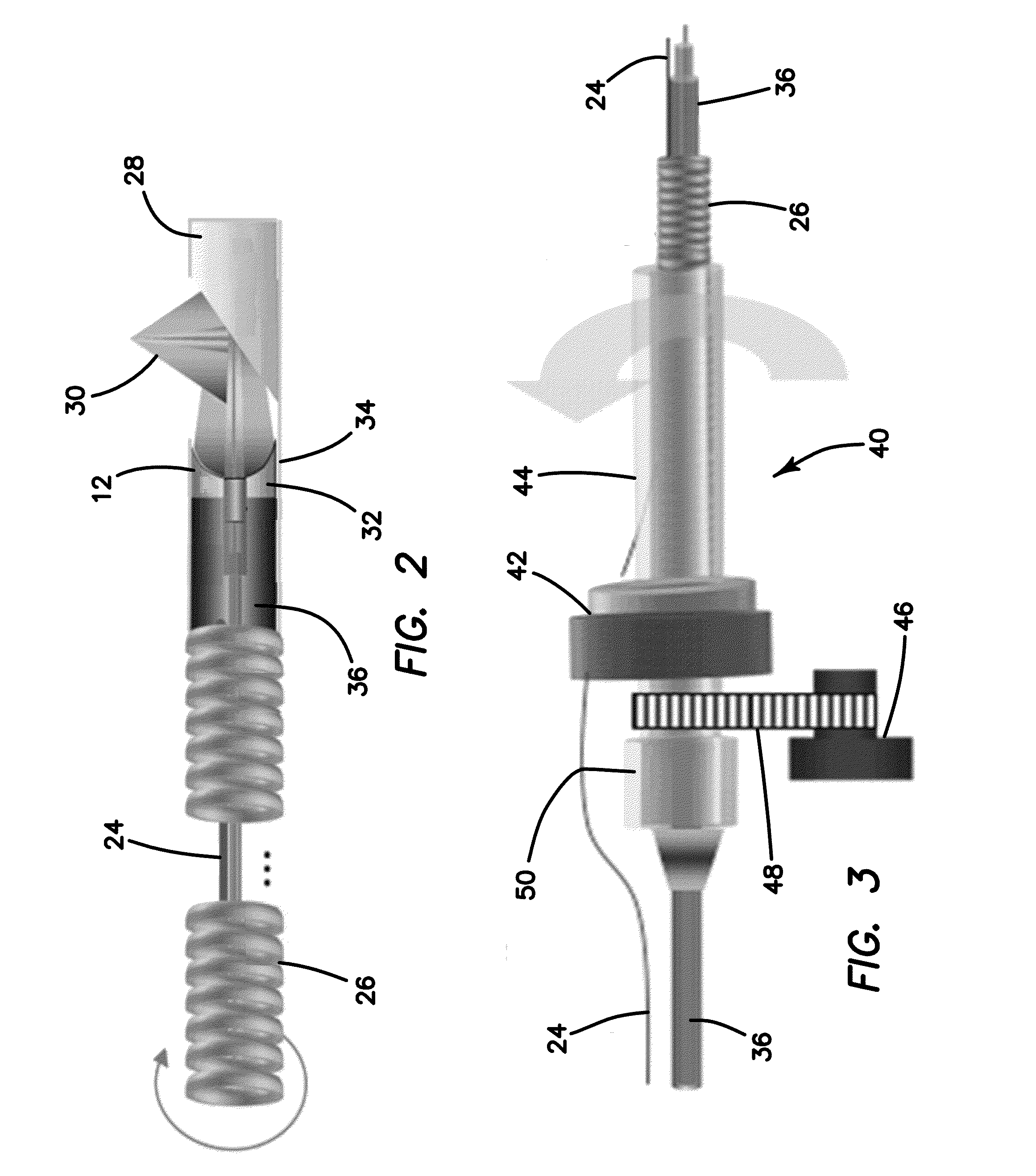
[0064]We have developed a PR-ARF-OCE system 10 that uses an acoustic wave generated acoustic radiation force to apply pressure to the tissue and uses phase-resolved OCT to evaluate the elastic properties of vascular tissue. The ARF-OCE combines the high-speed excitation of ARF with sub-micrometer / nanometer detection sensitivity of phase resolved OCT. This elastography technique allows high-speed and high-resolution point-by-point mapping of local strains in atherosclerotic coronary tissue, which provide critical information to assess the vulnerability of plaques. Using a known excitation signal also allows us to sweep different frequencies and measuring the frequency response of tissues in order to detect the differences between normal and diseased tissue with altered elasticity by sensing resonance frequencies between tissues. Since the mechanical properties of fibrous and fatty plaques are different, PR-ARF-OCE also has the potential to differentiate between various plaque compone...


PUM


Abstract

A method of using an integrated intraluminal imaging system includes an optical coherence tomography interferometer (OCT), an ultrasound subsystem (US) and a phase resolved acoustic radiation force optical coherence elastography subsystem (PR-RAF-OCE). The steps include performing OCT to generate a returned optical signal, performing US imaging to generate a returned ultrasound signal, performing PR-ARF-OCE to generate a returned PR-ARF-OCE signal by generating a amplitude modulated ultrasound beam or chirped amplitude modulated ultrasound beam to frequency sweep the acoustic radiation force, measuring the ARF induced tissue displacement using phase resolved OCT method, and the frequency dependence of the PR-ARF-OCE signal, processing the returned optical signal, the returned ultrasound signal and the measured frequency dependence of the returned PR-ARF-OCE optical coherence elastographic signal to quantitatively measure the mechanical properties of the identified tissues with both spectral and spatial resolution using enhanced materials response at mechanically resonant frequencies to distinguish tissues with varying stiffness, to identify tissues with different biomechanical properties and to measure structural and mechanical properties simultaneously.

Description

RELATED APPLICATIONS[0001]The present application is related to U.S. Provisional Patent Application, Ser. No. 61 / 783,178, filed on Mar. 14, 2013, which is incorporated herein by reference and to which priority is claimed pursuant to 35 USC 119.GOVERNMENT RIGHTS[0002]This invention was made with government support under contract numbers R01EB-10090, R01EY-021519, R01HL-105215, R01HL-103764, and P41EB-015890 awarded by the National Institutes of Health, and Air Force Office of Scientific Research (FA9550-04-0101). The government has certain rights in the invention.BACKGROUND[0003]1. Field of the Technology[0004]The disclosure relates to the field of medical device, multimodality intravascular imaging systems, multimodality endoscopic imaging systems, optical coherence elastography, and acoustic radiation force systems.[0005]2. Description of the Prior Art[0006]Atherosclerosis is a complex disease in which multiple plaques build up within the arteries. The major cause of cardiovascular...

Claims


Application Information

Patent Timeline
Patent Type & Authority Applications(United States)
IPC IPC(8): A61B8/08A61B5/00A61B8/12A61B8/00
CPCA61B8/485A61B8/4416A61B8/5261A61B8/12A61B5/0066A61B5/0035A61B5/02007A61B8/445
Inventor CHEN, ZHONGPINGQI, WENJUANLI, RUI
Owner RGT UNIV OF CALIFORNIA
Why Eureka
Try Eureka