Method for performing an endoscopic mucosal resection

a mucosal resection and endoscopic technology, applied in the field of medical devices and methods, can solve the problems of difficult to remove the entire suspect region, risk of penetrating the muscularis during cutting,

Active Publication Date: 2007-11-01
ETHICON ENDO SURGERY INC
View PDF23 Cites 10 Cited by
  • Summary
  • Abstract
  • Description
  • Claims
  • Application Information

AI Technical Summary

Problems solved by technology

However, such techniques have presented several disadvantages, including the difficulty associated

Method used


Image

  • Method for performing an endoscopic mucosal resection
  • Method for performing an endoscopic mucosal resection
  • Method for performing an endoscopic mucosal resection

Examples


Embodiment Construction

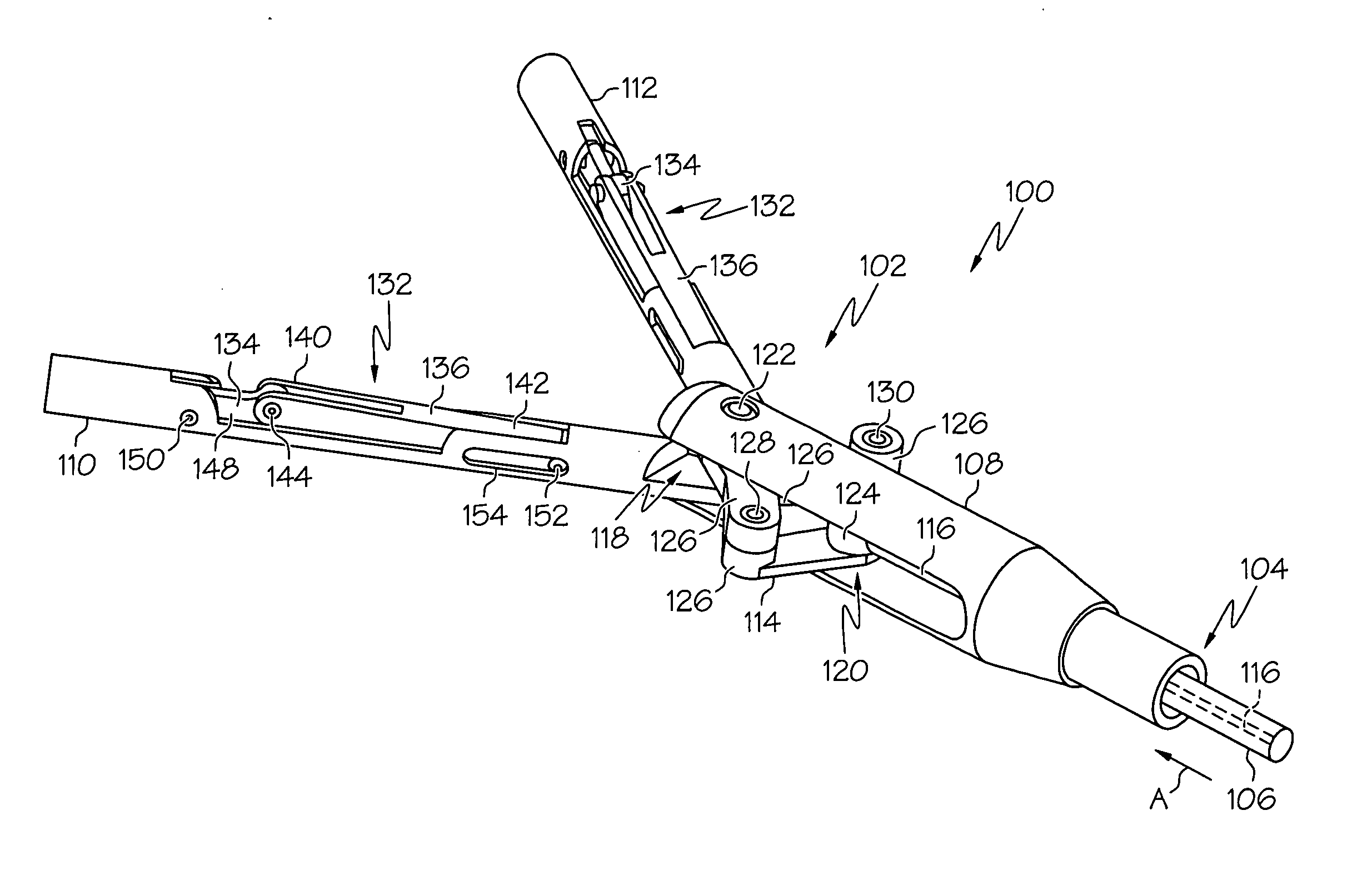
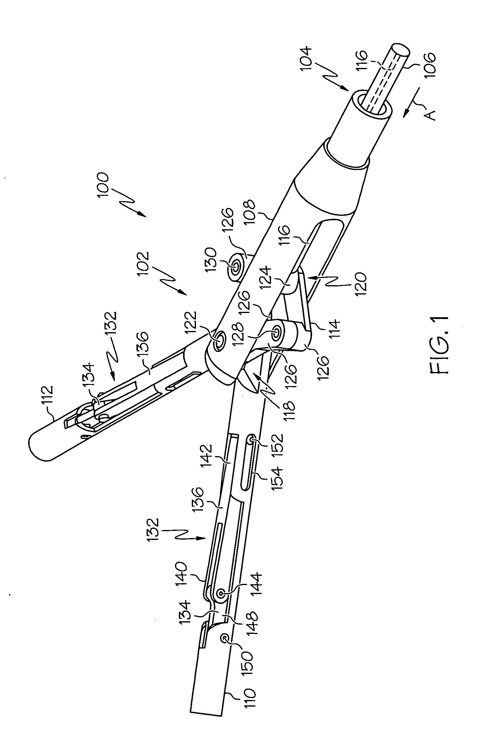
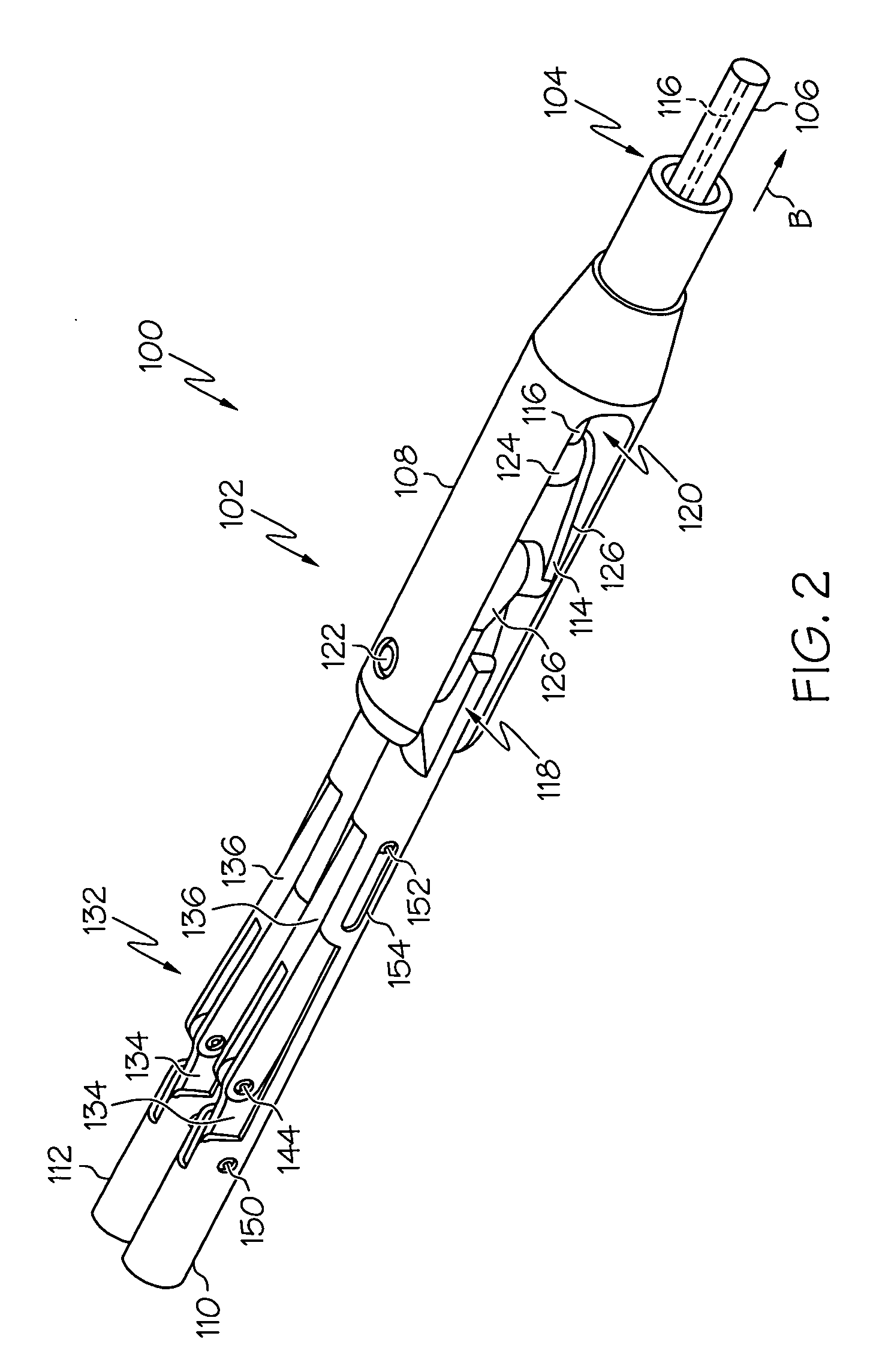
[0033] Referring to FIGS. 1-4, a first aspect of an improved EMR device, generally designated 100, may include a dissection / actuation assembly 102 disposed on the distal end 104 of an elongated shaft 106. The assembly 102 and shaft 106 may be sized and shaped to be received through a natural orifice of the human body (not shown). The shaft 106 may be flexible and may have a length sufficient to navigate the human gastrointestinal tract during an endoscopic procedure.

[0034] The assembly 102 may include a head 108, a first moveable arm 110, a second moveable arm 112, a linkage assembly 114 and an actuation link or cable 116. The linkage assembly 114 may be comprised of four pivotally connected links 126 and may be disposed within the head 108. The actuation cable 116 may extend through the elongated shaft 106 such that it is accessible by a user.

[0035] The linkage assembly 114 may include a distal end 118 and a proximal end 120, wherein the distal end 118 may be connected to the fir...


PUM


Abstract

A method for resecting tissue from an organ of a patient, the organ being comprised of at least a first layer, a second layer and a third layer, the method including the steps of providing an EMR device with access to the organ, the EMR device including at least one deployable cutting element disposed on at least one moveable arm, identifying a target tissue area for resection, forming an incision through the first and second layers without penetrating the third layer, positioning the EMR device through the incision such that the device is positioned between the first third layers, advancing the moveable arm between the first and third layers, deploying the cutting element such that the cutting element is advanced in the direction of the first layer and away from the third layer and, while the EMR device is positioned between the first and third layers, cutting through the first layer.

Description

FIELD OF THE INVENTION [0001] The present application relates to medical devices and methods and, more particularly, to medical devices and methods for performing resection procedures within the gastrointestinal and esophageal passages of the human body. BACKGROUND OF THE INVENTION [0002] Gastric cancers in the human body often stem from precursor lesions and polyps that develop into superficial tumors or other growths. Such lesions and growths typically begin in the mucosal layer of the gastrointestinal tract (e.g., the mucosa of the colon) and, as the cancer develops, may spread to the submucosal layer and beyond. Therefore, many physicians agree that successful cancer treatment and prevention typically requires the identification and removal of suspect tissue from the gastrointestinal tract. [0003] Accordingly, physicians may collect samples of tissue from the gastrointestinal tract (e.g., samples of the mucosa) and test the sample tissue for the presence of cancerous cells. The ...

Claims


Application Information

Patent Timeline
IPC IPC(8): A61B18/14
CPCA61B18/04A61B18/082A61B18/1445A61B18/1492A61B2017/00269A61B2018/144A61B2018/00482A61B2018/1253A61B2018/1407A61B2018/1432A61B2017/2945
Inventor NOBIS, RUDOLPH H.LU, IFUNG
Owner ETHICON ENDO SURGERY INC
Why Eureka
Try Eureka