Tissue Resection Device

a resection device and mucosal technology, applied in the field of mucosal resection devices, can solve the problems of large flat mucosal lesions removed in a single operation, numerous problems for current endoscopic tools and techniques, and inability to perform full thickness biopsies

Active Publication Date: 2011-04-14
APOLLO ENDOSURGERY INC
View PDF8 Cites 8 Cited by
  • Summary
  • Abstract
  • Description
  • Claims
  • Application Information

AI Technical Summary

Benefits of technology

[0016]In accordance with one aspect of the present invention there is provided a mucosal layer tissue resection device for removing a target area of mucosal tissue in the digestive tract of a mammal. The mucosal layer tissue resection device includes a tubular shaft having proximal and distal ends and a lumen extending therethrough. Located at the distal end of the shaft are first and second elongated tubular tissue guides which are formed of a nonconductive material and biased outwardly. Each tubular tissue guide has an elongated slot through the wall which extends longitudinally along at least a portion of the guide length. The slots of the tissue guides are positioned opposing each other. An electrosurgical loop cutter having an elongate electroconductive member slidably position

Problems solved by technology

While many of these devices and techniques have been useful in identifying and removing some neoplastic lesions of the mucosal layer as well as providing access to general locations within the digestive tract for the placement of submucosal implants, there are some lesions and areas of the digestive tract which are extremely difficult to resect or access.
For example, the en bloc removal of large flat mucosal lesions presents numerous problems for current endoscopic tools and techniques.
Currently, access to these types of specimens requires full thickness biopsies which can be particularly difficult from an endoscopic approach requiring extremely skilled closure techniques.
While EMR techniques have been shown to be effective in treating some flat neoplastic lesions there are limitations and complications associated with these techniques.
A major limitation associated with this technique is the size of the lesion that can be resected.
While larger or irregular shaped lesions may be resected in a piecemeal fashion, this is undesirable since small portions of the lesion may remain.
Another limitation of these techniques includes uncertainty of the area being resected.
One complication associated with these EMR techniques is in relation to the use of t

Method used


Image

  • Tissue Resection Device
  • Tissue Resection Device
  • Tissue Resection Device

Examples


Embodiment Construction

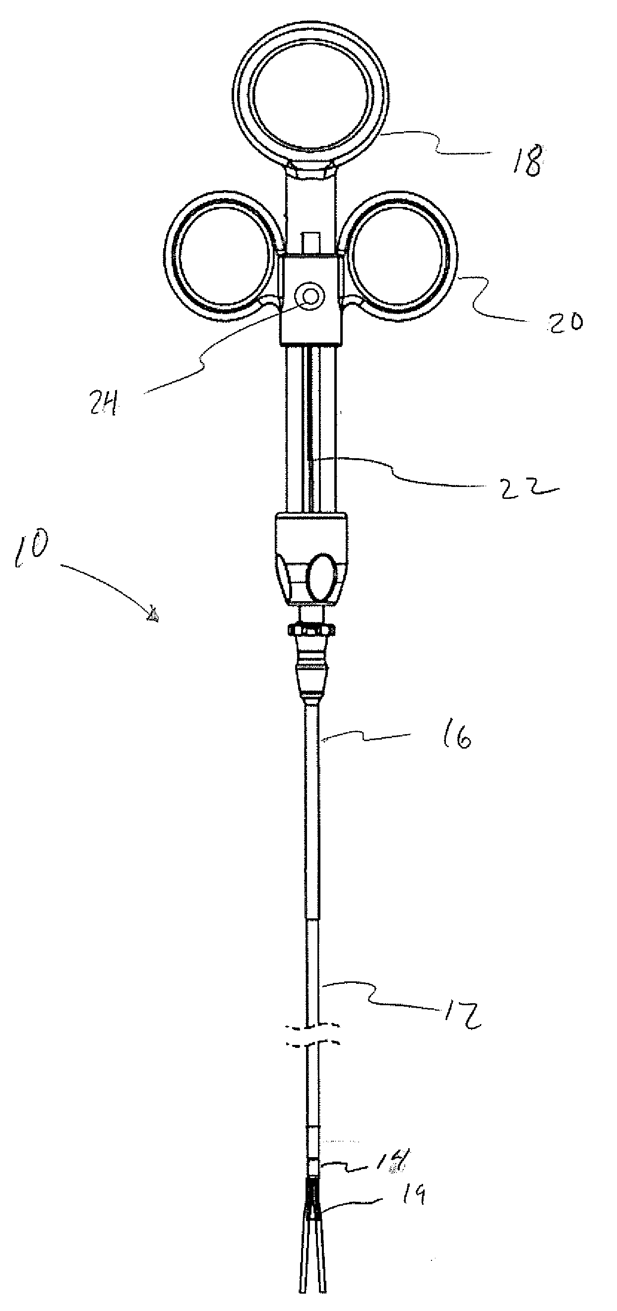
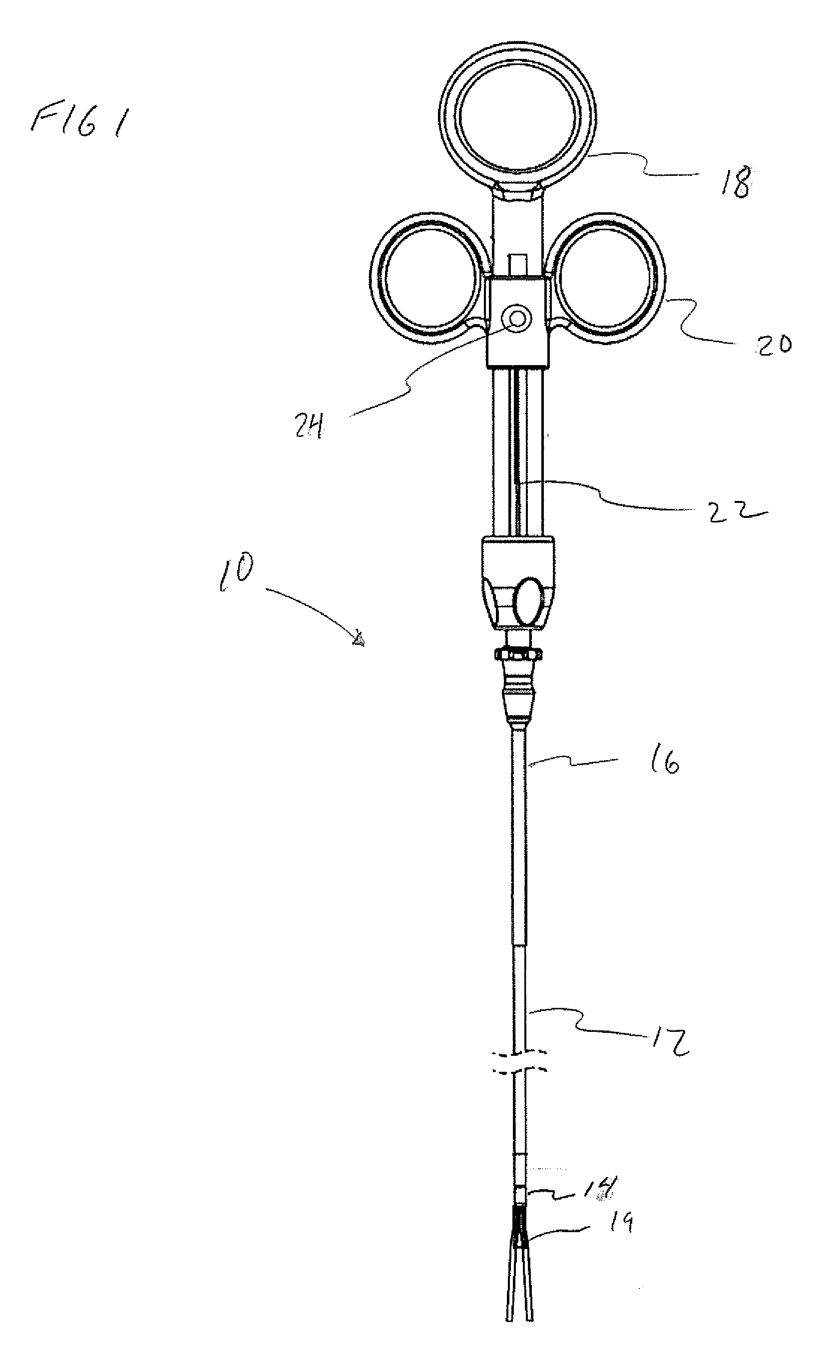
[0028]FIG. 1 illustrates a tissue resection device for performing a submucosal medical procedure to resect a portion of the mucosal layer in the digestive tract of a mammal according to an embodiment of the present invention. The tissue resection device 10 includes an elongate catheter 12 having a distal end 14, a proximal end 16 and a lumen extending therethrough. Handle assembly 18 is coupled to the proximal end 16 of catheter 12 and electrosurgical loop cutter wire 19 is positioned at the distal end. Handle assembly 18 includes a slide assembly 20 which is fixedly coupled to the proximal end of the cutting wire actuator 22 of the electrosurgical loop cutter wire 19. Slide assembly 20 includes an electrical connector 24 which is in electrical contact with proximal end 22 and facilitates the coupling of an electrosurgical generator to supply energy to cutter wire 19.

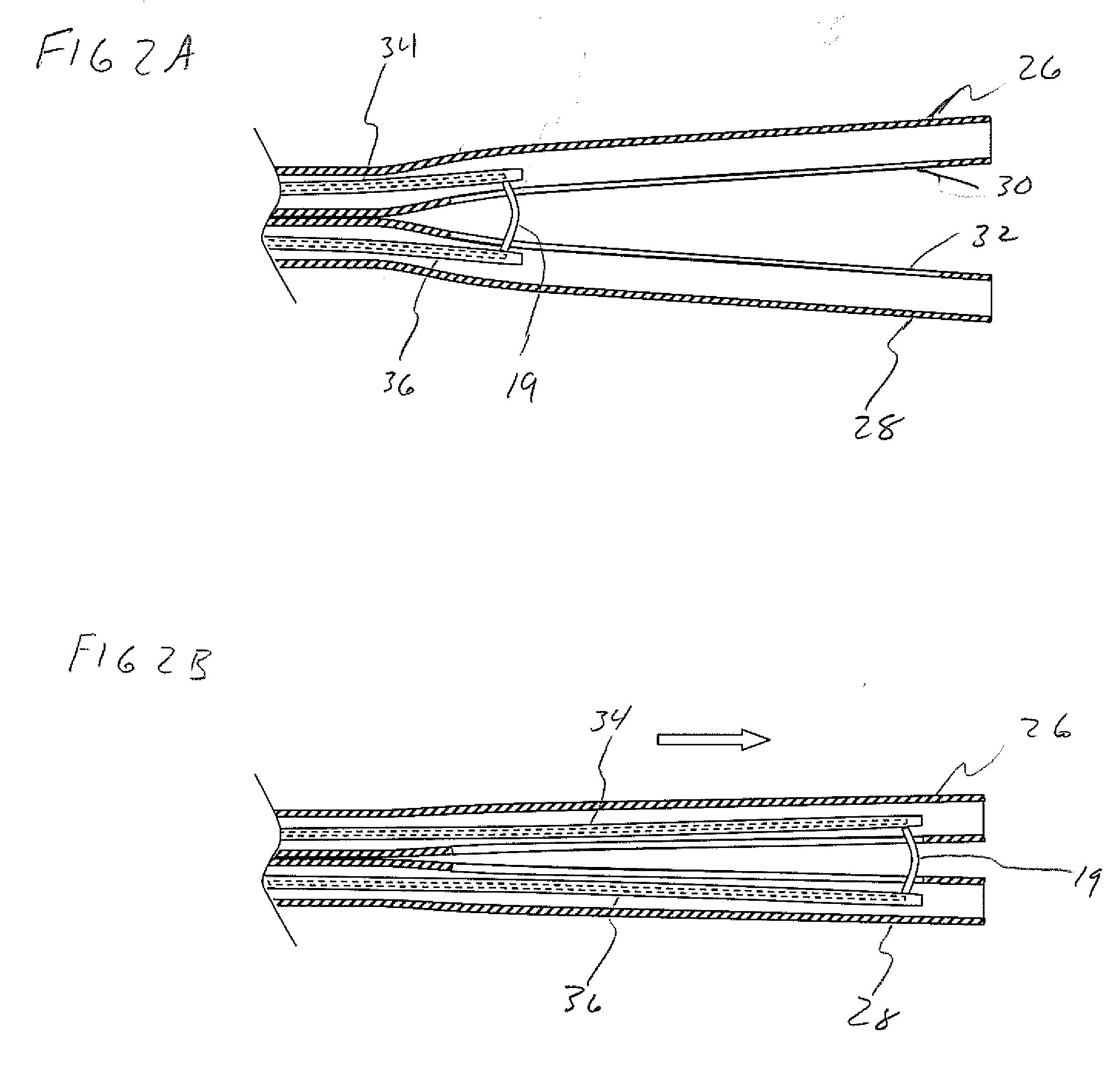
[0029]FIGS. 2A and 2B illustrate the distal end of tissue resection device 10 in more detail showing a partial cross ...


PUM


Abstract

Instruments and methods are provided for performing submucosal medical procedures in a desired area of the digestive tract using endoscopy. The instruments include a mucosal resection device, a tissue grasper and a snare. Systems include a combination of one or more of such instruments. Embodiments of various methods for performing the procedures are also provided.

Description

CROSS-REFERENCE TO RELATED APPLICATION[0001]This application claims benefit of U.S. Provisional Application No. 61 / 250,869, filed Oct. 12, 2009, which is hereby incorporated by reference herein in its entirety.FIELD OF THE INVENTION[0002]The present invention relates to a mucosal resection device and a method for performing submucosal medical procedures in a desired area of the digestive tract using an endoscope.BACKGROUND OF THE INVENTION[0003]The field of gastrointestinal endoscopy has for many years focused on diagnostic and therapeutic techniques to observe, modify and remove tissues located in the digestive tract. General endoscopic procedural techniques such as visualizing, dilating, cutting and manipulating tissue have been accomplished using flexible devices such as endoscopes, balloons, snares and electrosurgical tools well known in the art.[0004]While many of these devices and techniques have been useful in identifying and removing some neoplastic lesions of the mucosal la...

Claims


Application Information

Patent Timeline
IPC IPC(8): A61B18/00
CPCA61B18/14A61B2018/144A61B2018/1407A61B18/1445
Inventor MILLER, DAVIDDALTON, TODDGILKEY, MITCH
Owner APOLLO ENDOSURGERY INC
Why Eureka
Try Eureka