

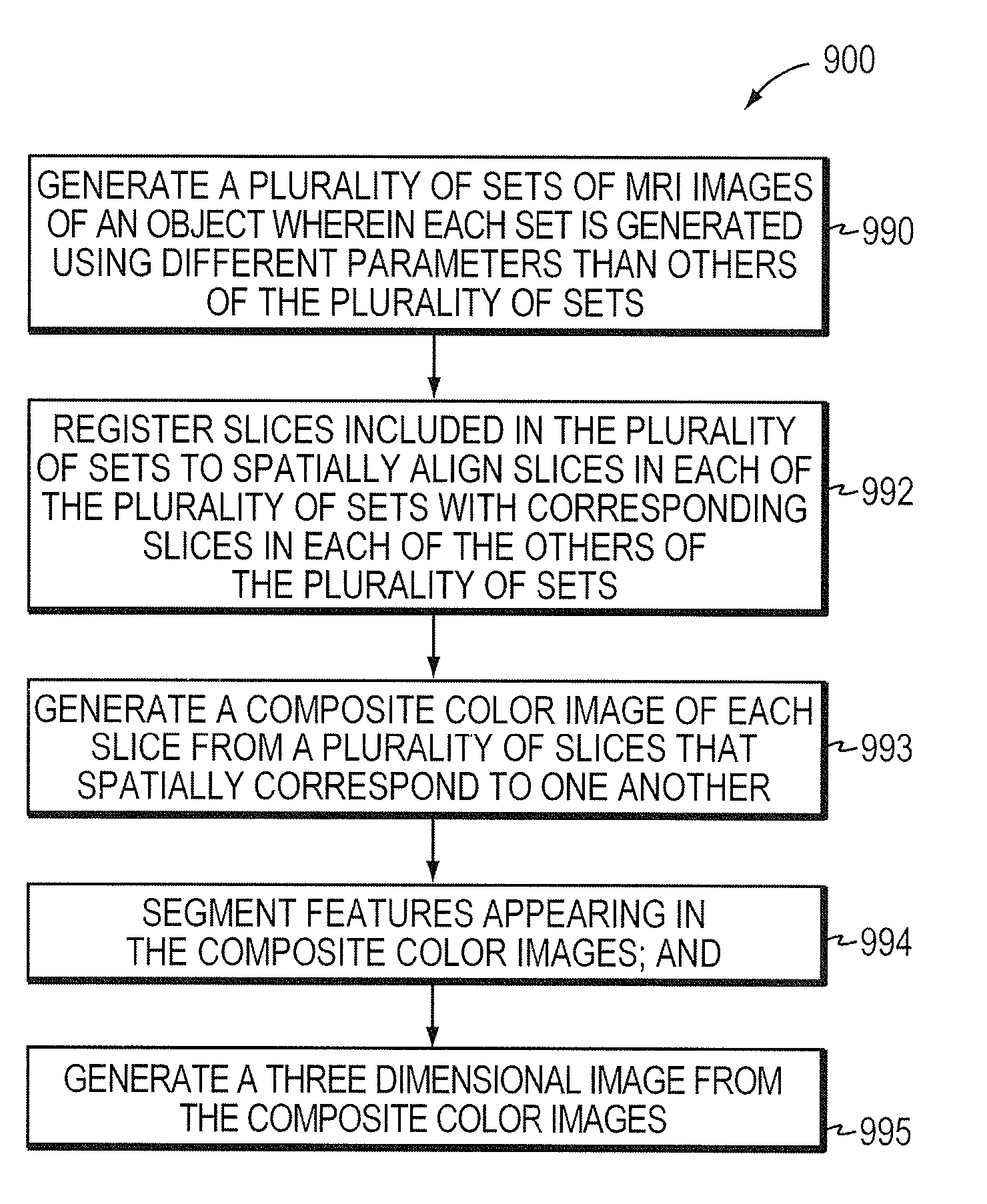
Three-dimensional rendering of MRI results using automatic segmentation
a three-dimensional rendering and image technology, applied in image analysis, medical science, diagnostics and other directions, can solve the problems of nuanced diagnosis and analysis performed using mri images, considerable experience, and difficult and time-consuming for a professional, and achieves a high degree of confidence. the effect of accurate diagnosis and analysis
- Summary
- Abstract
- Description
- Claims
- Application Information
AI Technical Summary
Benefits of technology
Problems solved by technology
Method used
Image

Examples
Embodiment Construction
[0028]This invention is not limited in its application to the details of construction and the arrangement of components set forth in the following description or illustrated in the drawings. The invention is capable of other embodiments and of being practiced or of being carried out in various ways. Also, the phraseology and terminology used herein is for the purpose of description and should not be regarded as limiting. The use of “including,”“comprising,” or “having,”“containing,”“involving,” and variations thereof herein, is meant to encompass the items listed thereafter and equivalents thereof as well as additional items.
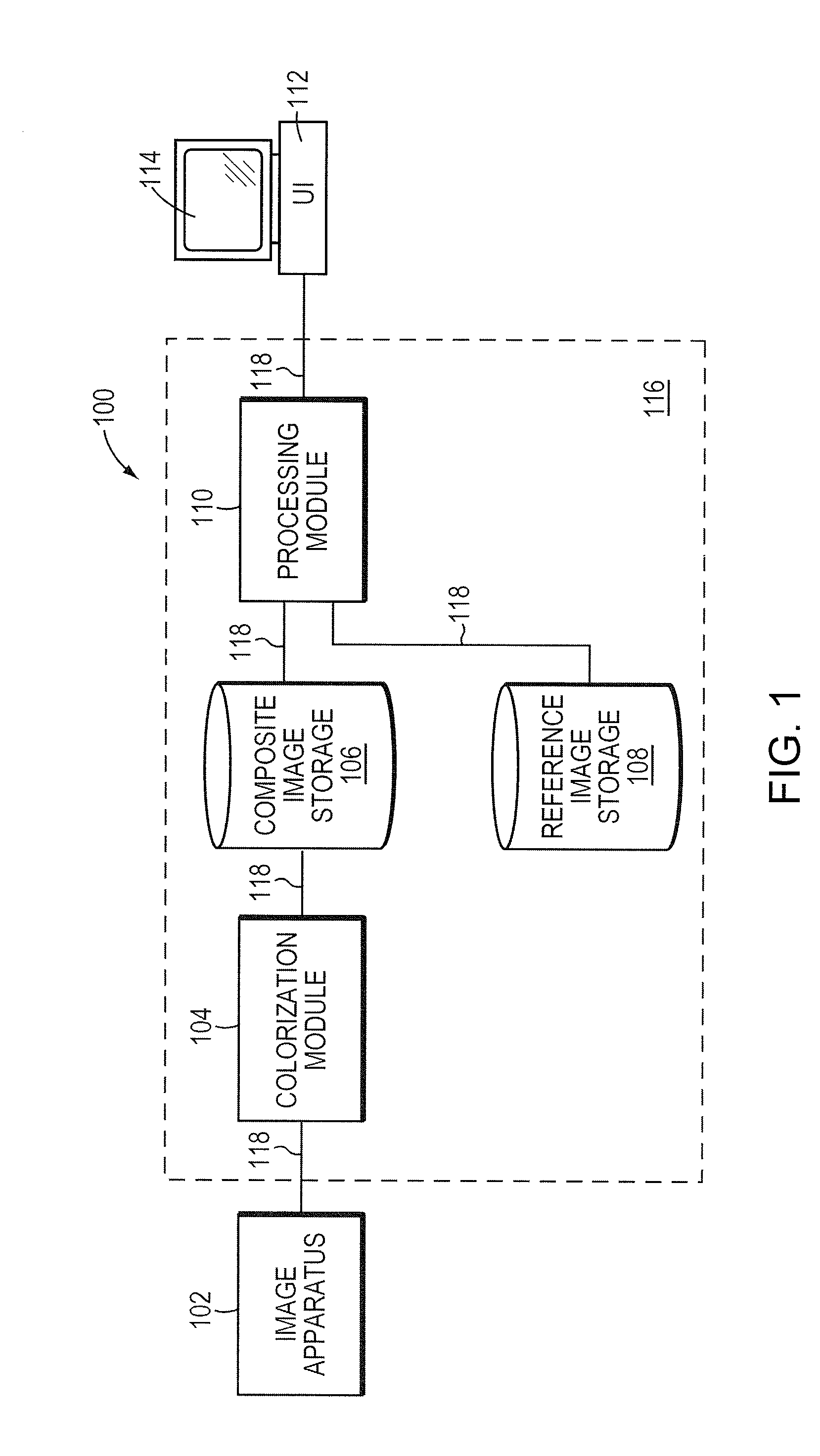
[0029]Referring to FIG. 1, a system for processing color MRI images for diagnostic analysis is illustrated. The system 100 includes image generation apparatus 102, colorization module 104, a composite image storage module 106, a reference image storage module 108, a processing module 110 and a user interface 112. The image generation apparatus 102 may be any of ...
PUM
 Abstract
Description
Claims
Application Information