X-ray reference device and method of use

a reference device and x-ray technology, applied in the field of radiographic reference tools, can solve the problems of high cost of devices, difficult to determine the size needed from digital radiographs, and inability to tell the dentist from radiographs the length of teeth or the amount of bone present,

Inactive Publication Date: 2008-02-07
MUCKLER MICHAEL P
View PDF16 Cites 11 Cited by
  • Summary
  • Abstract
  • Description
  • Claims
  • Application Information

AI Technical Summary

Problems solved by technology

This is fine for diagnosing caries, but when these images are taken in preparation for the placement of a dental implant or root canal treatment the dentist can no longer tell from the radiograph the length of the tooth or the amount of bone present.
Therefore if implants are necessary the sizes needed are hard to determine from a digital radiograph.
The problem here is that these devices are costly.

Method used


Image

  • X-ray reference device and method of use
  • X-ray reference device and method of use
  • X-ray reference device and method of use

Examples


Embodiment Construction

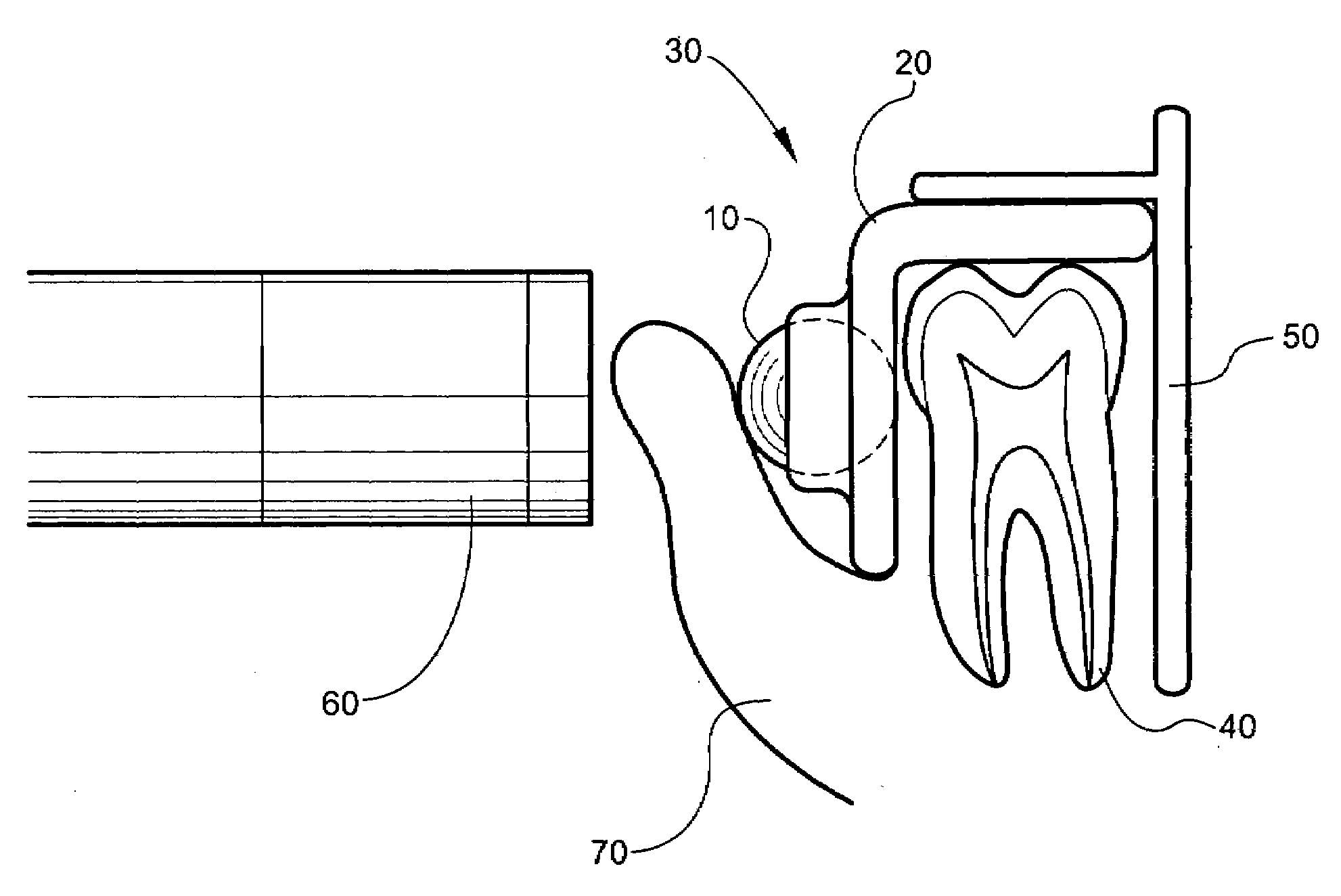
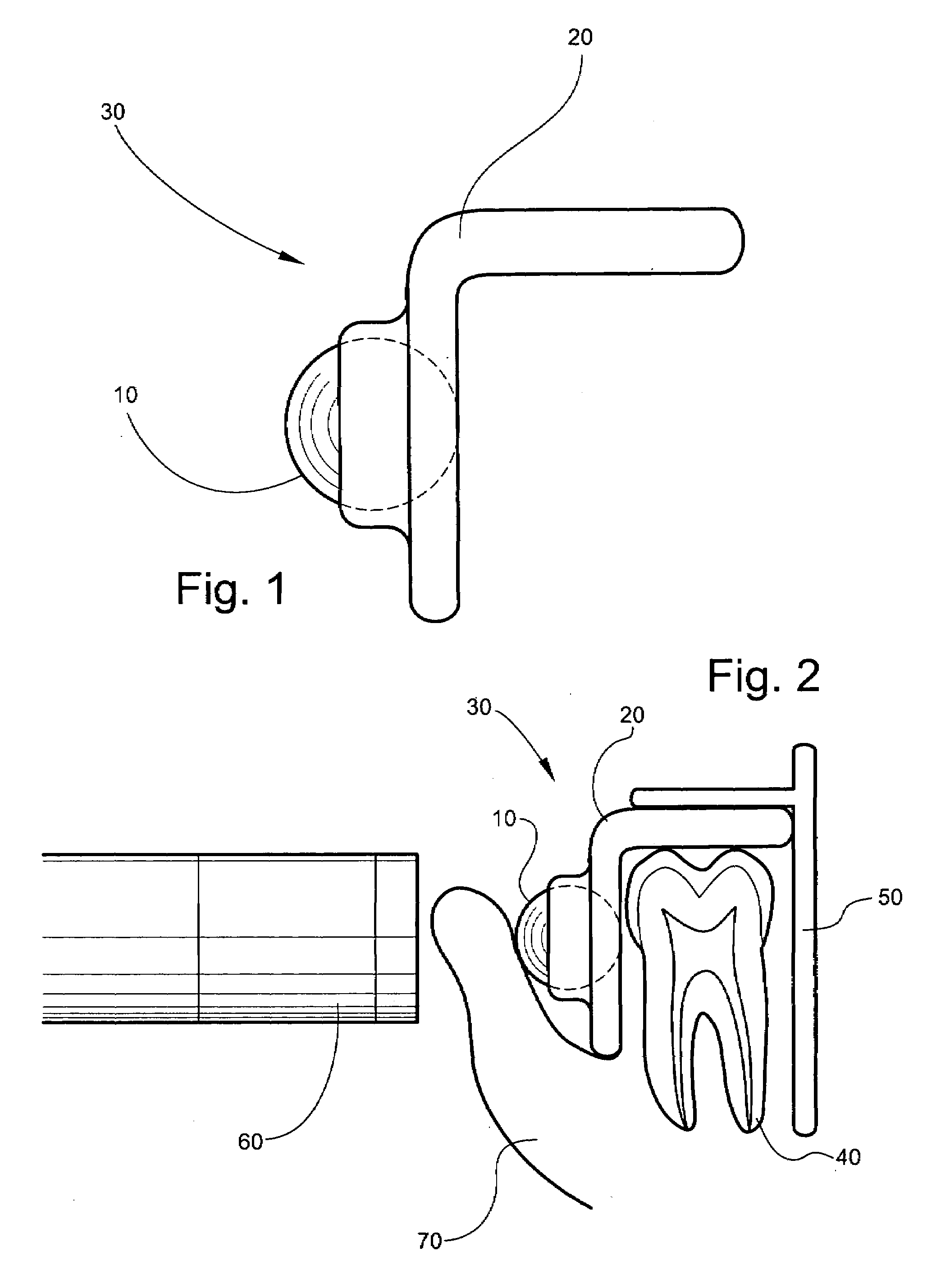
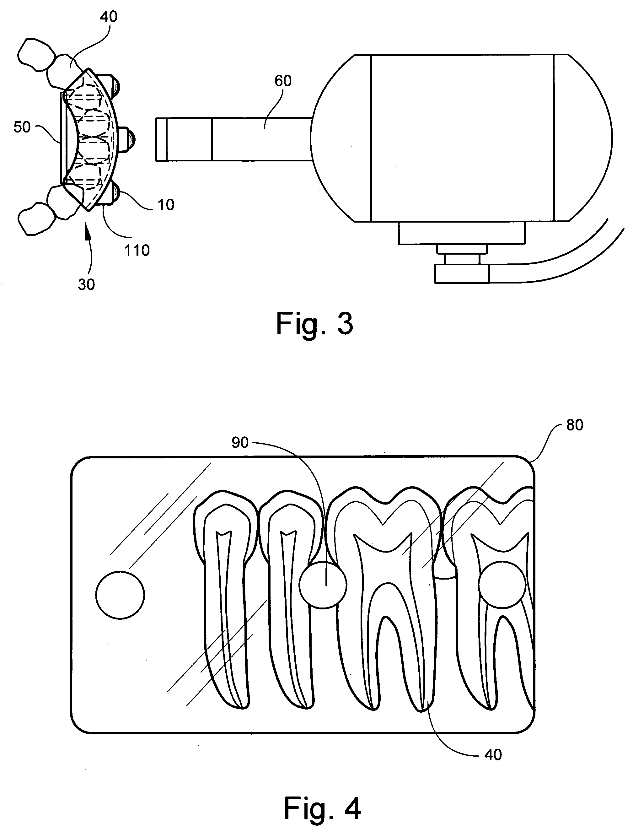
[0016]The X-ray reference device 30, FIG. 1, is a measuring tool that allows the dentist to accurately measure the length of teeth for root canal treatments and measure the amount of bone height present for implant placement. This X-ray reference device 30 comes in several sizes for the anterior and posterior arch segments. The X-ray reference device 30, can be either a plastic, rubber, or cardboard instrument. The X-ray reference device 30, may containing one radiopaque marker 12, FIG. 8, or multiple radiopaque markers 14, 16 and 18, FIG. 6. The radiopaque marker 10 is a reference object of a known size which is capable of leaving an image, a reference shadow 90, FIG. 4, on periapical radiographs 80 or panoramic images, 100, FIG. 9. The radiopaque marker 10 may be spherical or another geometric shape of a known size. When the X-ray reference device 30 is placed on a facial surface of a tooth 40, FIG. 2, it provides a reference shadow 90, FIG. 4, in taking periapical radiographs 80 ...


PUM


Abstract

A dental X-ray reference device comprised of a radiopaque marker having a known dimension and a tooth overlay molding adapted to secure the radiopaque marker in close proximity to a tooth. Also disclosed is a process for measuring teeth for implant placement or root canal treatment. This process comprises the steps of providing a dental X-ray reference device comprised of a radiopaque marker having a known dimension and a tooth overlay molding adapted to secure the radiopaque marker in close proximity of a tooth, or alveolar ridge. The dental X-ray reference device is then positioned in a mouth of a patient. The radiopaque marker is positioned over the edentulous area or over a tooth. Then an X-ray sensor is positioned behind the tooth, or alveolar ridge, and behind the X-ray reference device. A radiograph of the tooth or alveolar ridge, and the radiopaque marker is taken.

Description

BACKGROUND OF THE INVENTION[0001]The present invention provides a radiography reference tool, which provides a known reference for use with digital X-ray equipment to facilitate dental work such as root canal treatment and the placement of dental implants.[0002]Radiographs are used extensively by dentists to look for damage or decay to teeth. Traditionally radiographs where taken with film which was placed directly into the mouth and then exposed with a beam emitter from an X-ray gun. This would take an image which is roughly the same size as the tooth, but it would require that the film be processed so that it can be read which takes time. Therefore many dentists have gone to using digital radiographs where the images are available immediately after being taken and the images are usually displayed larger than actual-size to better read the detail of the image. This is fine for diagnosing caries, but when these images are taken in preparation for the placement of a dental implant or...

Claims


Application Information

Patent Timeline
Patent Type & Authority Applications(United States)
IPC IPC(8): A61C3/00
CPCG03B42/042
Inventor MUCKLER, MICHAEL P.
Owner MUCKLER MICHAEL P
Why Eureka
Try Eureka