
Digital radiography apparatus

a radiography apparatus and digital technology, applied in electrical apparatus, medical science, diagnostics, etc., can solve the problems of mechanical complexity, disadvantages in usability, cost and reliability, etc., and achieve the effects of cost and reliability, suitable positioning, and usability

Inactive Publication Date: 2008-03-20
CARESTREAM HEALTH INC
View PDF8 Cites 16 Cited by
  • Summary
  • Abstract
  • Description
  • Claims
  • Application Information

AI Technical Summary

Benefits of technology

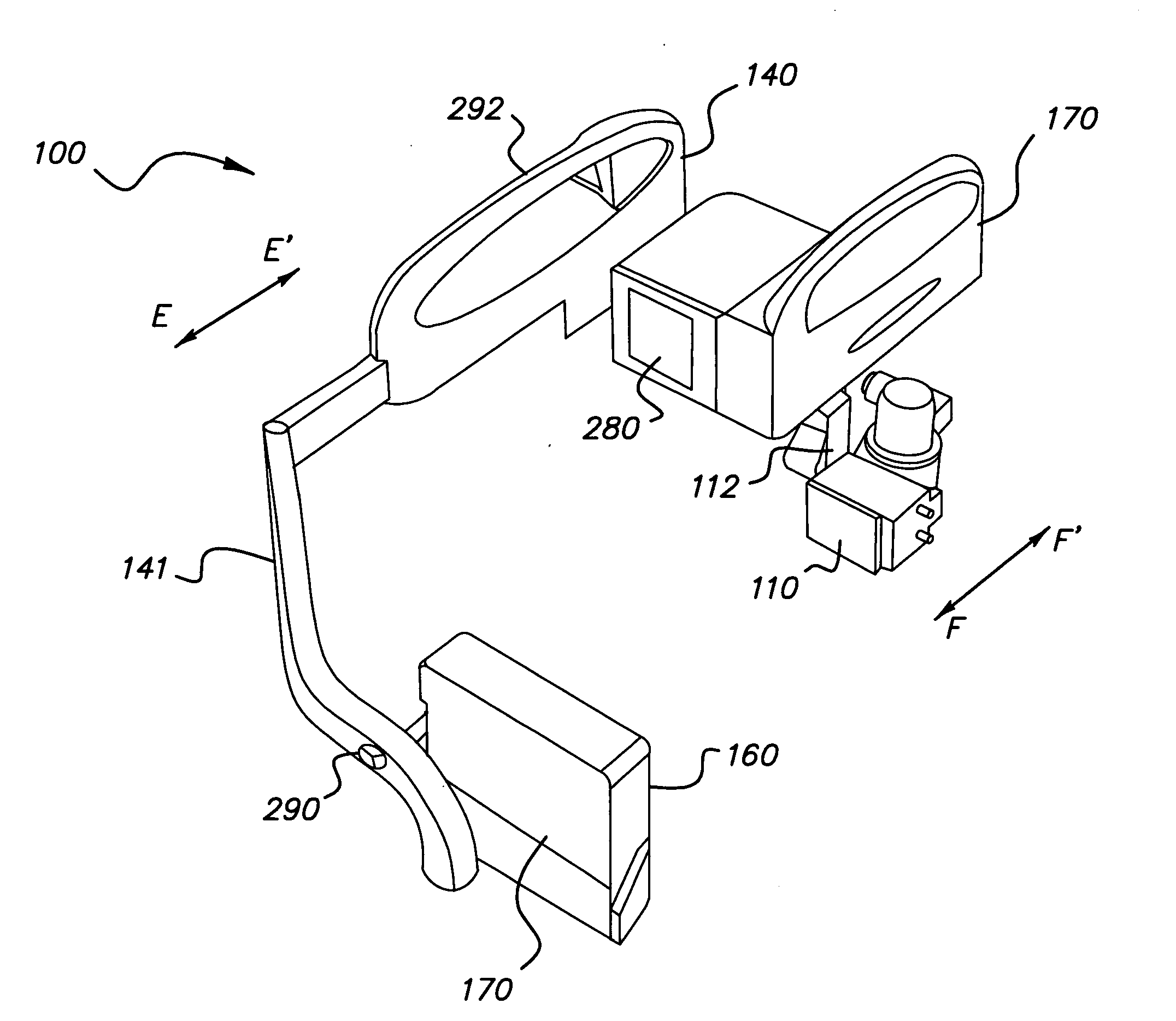
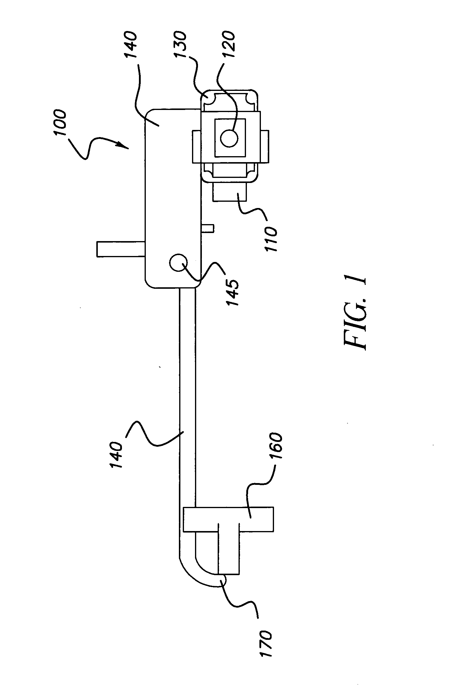
[0011]The present invention provides a compact, adjustable digital radiography imaging system where the X-ray source and X-ray imaging detector are coupled. Typical imaging systems are generally much larger, or have separate pieces of equipment that work together; such systems are mechanically complex, and have disadvantages in usability, cost and reliability.
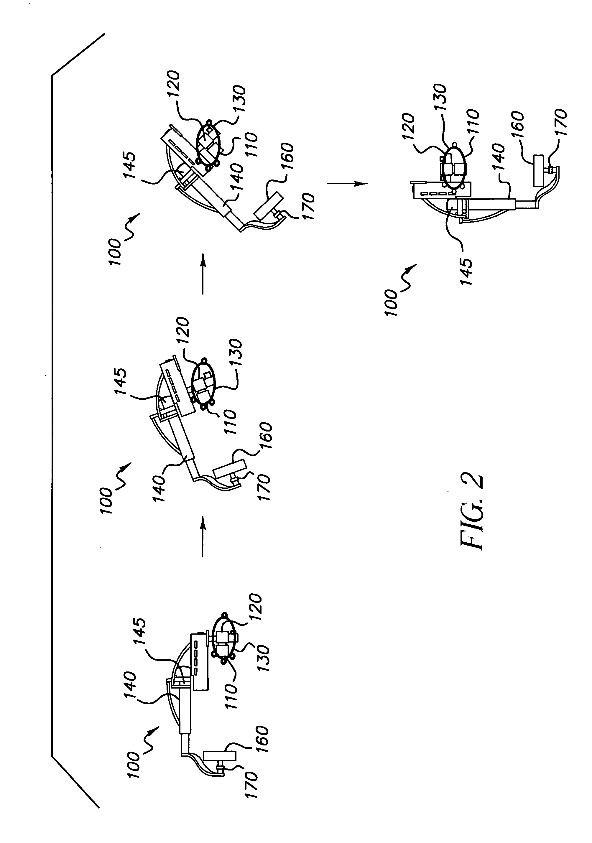
[0012]The adjustability of the present invention allows an operator to position the X-ray source and X-ray imaging detector to achieve suitable positioning to accommodate subjects for imaging (including ambulatory and non-ambulatory patients standing, reclining or in seated position).

Problems solved by technology

Typical imaging systems are generally much larger, or have separate pieces of equipment that work together; such systems are mechanically complex, and have disadvantages in usability, cost and reliability.

Method used


Image

  • Digital radiography apparatus
  • Digital radiography apparatus
  • Digital radiography apparatus

Examples


Embodiment Construction

[0037]The following is a detailed description of the preferred embodiments of the invention, reference being made to the drawings in which the same reference numerals identify the same elements of structure in each of the several figures.

[0038]The present invention is directed to a digital radiography system wherein an X-ray or other suitable radiation source projects radiation through a subject (e.g., patient) to produce an image captured by an imaging detector. The radiation source and imaging detector can be positioned in various orientations to capture an image of a patient. The present invention provides multiple redundant work zones, each work zone including appropriate setup controls and a display for setup and operation of the digital radiography system. The description that follows describes an embodiment using X-ray imaging; however, it is noted that the apparatus and method of the present invention can be more applied for other suitable types of diagnostic imaging.

[0039]R...


PUM


Abstract

A radiography apparatus having an x-ray source, an x-ray imaging detector, and a support structure. The support structure is coupled to the x-ray source and to the x-ray detector and rotatable about two orthogonal axes and linearly moveable. The x-ray source and the x-ray imaging detector are each independently rotatable about third and fourth axes, respectively. The apparatus provides an operator with a number of degrees of freedom of motion of the x-ray source and the x-ray imaging detector to move them to different positions relative to a subject.

Description

RELATED APPLICATIONS[0001]Reference is made to commonly assigned application entitled “Digital Radiography Imaging System with Rotatable Display and Controls”, U.S. Ser. No. ______ (Kodak Docket No. 91550), filed on even date, in the names of Muszak et al., incorporated herein by reference.[0002]Reference is made to commonly assigned patent application entitled “Radiography Apparatus with Multiple Work Zones”, U.S. Ser. No. ______ (Kodak Docket No. 92497), filed on even date, in the name of Muszak et al., incorporated herein by reference.FIELD OF THE INVENTION[0003]This invention generally relates to digital radiography and more particularly relates to a digital radiography imaging apparatus.BACKGROUND OF THE INVENTION[0004]Digital Radiography (DR) systems are employed in the field of medical technology for capturing digital medical images. Some digital radiography imaging systems have an X-ray source and an X-ray imaging detector that are coupled together and supported in a manner ...

Claims


Application Information

Patent Timeline
Patent Type & Authority Applications(United States)
IPC IPC(8): H05G1/02
CPCA61B6/4464A61B6/548A61B6/587
Inventor CHAPMAN, RAYMOND P.
Owner CARESTREAM HEALTH INC
Learn More