Radiography apparatus with multiple work zones

a radiography apparatus and work zone technology, applied in electrical apparatus, medical science, diagnostics, etc., can solve the problems of mechanical complexity, disadvantages in usability, cost and reliability, etc., and achieve the effects of reducing the difficulty of adjusting the position, and improving the usability and reliability

Inactive Publication Date: 2008-03-20
CARESTREAM HEALTH INC
View PDF8 Cites 31 Cited by
  • Summary
  • Abstract
  • Description
  • Claims
  • Application Information

AI Technical Summary

Benefits of technology

[0012]The present invention provides a compact, adjustable digital radiography imaging system where the X-ray source and X-ray imaging detector can be positioned in positions achievable with conventional floor mounted systems, with the additional feature of providing a display and controls which have a given orientation with respect to the operator. Typical imaging systems are generally much larger, or have separate pieces of equipment that work together. Such systems are mechanically complex, and have disadvantages in usability, cost and reliability.
[0013]The adjustability of the present invention allows an operator to position the X-ray source and X-ray imaging detector to achieve suitable positioning to accommodate subjects for imaging (including ambulatory and non-ambulatory patients standing, reclining or in seated position), and provides a display and controls which retain the same orientation with respect to the operator. Thus, the operator control interface and display of the present invention is accessible to the operator so that the position of the operator does not have to change when the position of the X-ray source and X-ray imaging detector are changed.
[0014]The position of the operating control interface and its display relative to the operator is relevant for critical environments, such as emergency or trauma rooms. Advantages of the operator control interface include, but are not limited to, greater legibility and readability of the display, fewer errors made by an operator in orienting the system to procure images, and other related advantages. By virtue of its size and placement of the operator control interface relative to the operator, this invention minimizes the potential for injury to an operator or patient by accidental contact with the hardware. The invention can reduce the potential for collision between with obstructions in the installation environment by providing the operator with familiar controls.

Problems solved by technology

Such systems are mechanically complex, and have disadvantages in usability, cost and reliability.

Method used


Image

  • Radiography apparatus with multiple work zones
  • Radiography apparatus with multiple work zones
  • Radiography apparatus with multiple work zones

Examples


Embodiment Construction

[0033]The following is a detailed description of the preferred embodiments of the invention, reference being made to the drawings in which the same reference numerals identify the same elements of structure in each of the several figures.

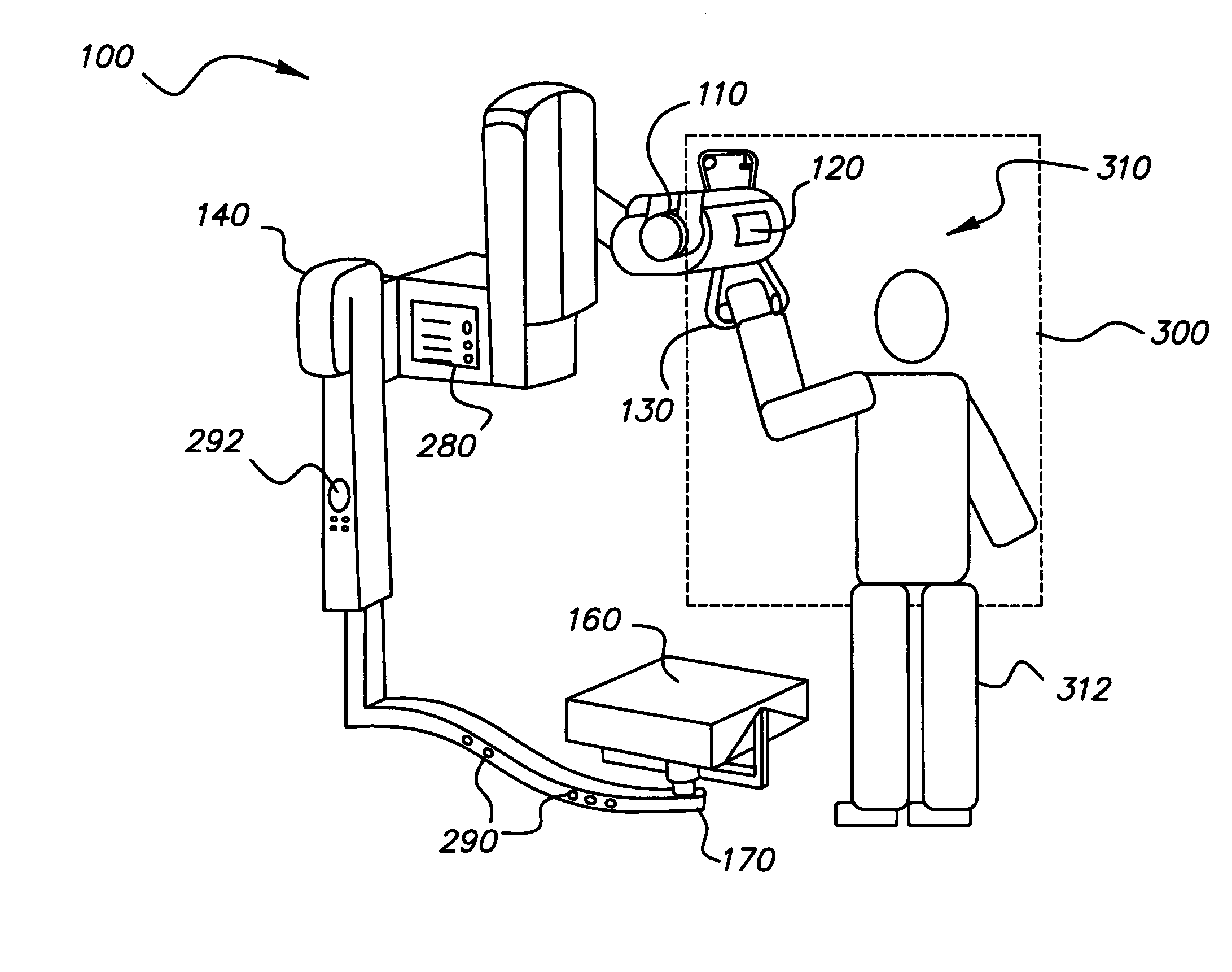
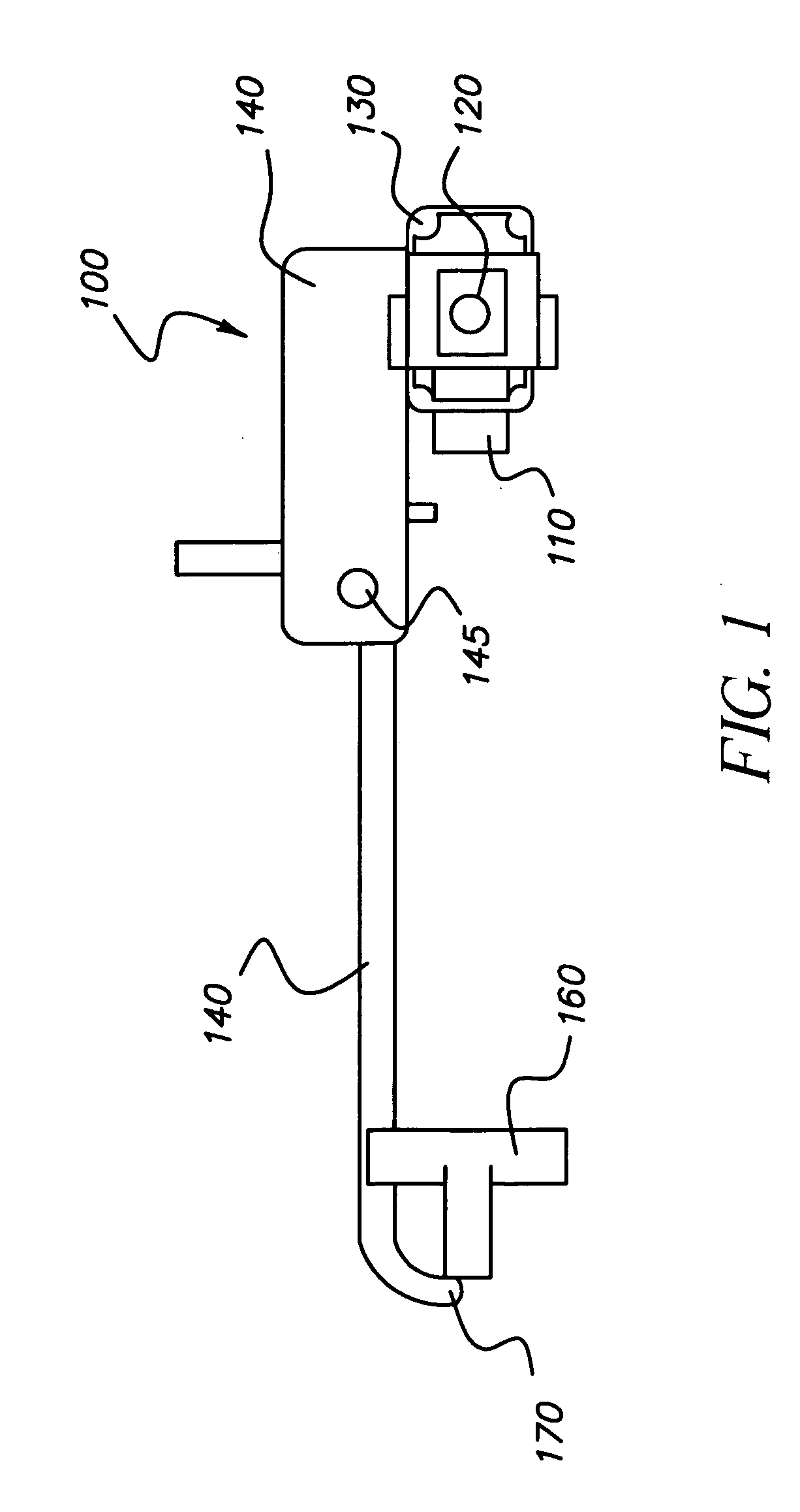
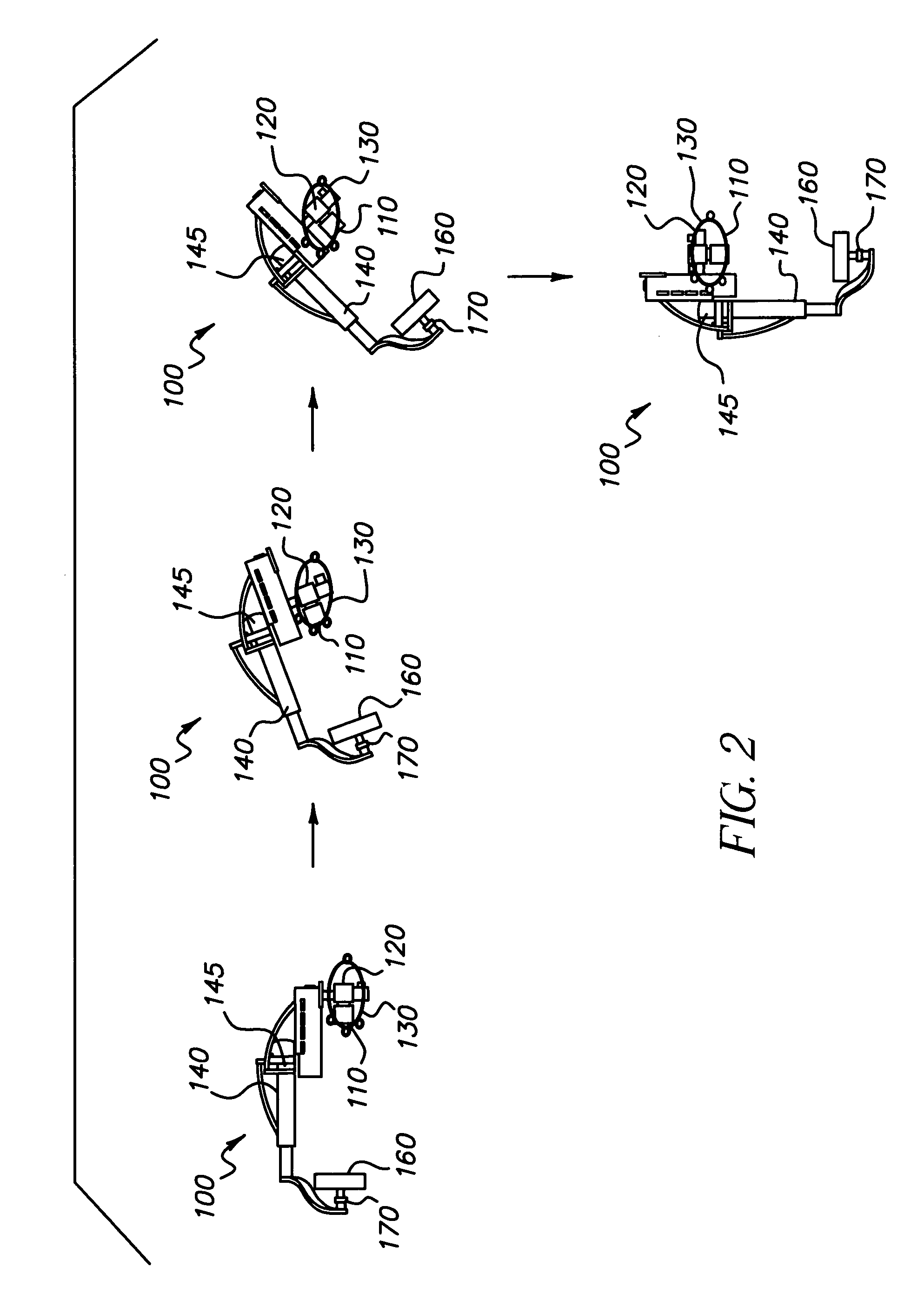
[0034]The present invention is directed to a digital radiography system wherein an X-ray or other suitable radiation source projects radiation through a subject (e.g., patient) to produce an image captured by an imaging detector. The radiation source and imaging detector can be positioned in various orientations to capture an image of a patient. The present invention provides multiple redundant work zones, each work zone including appropriate setup controls and a display for setup and operation of the digital radiography system. The description that follows describes an embodiment using X-ray imaging; however, it is noted that the apparatus and method of the present invention can be more applied for other suitable types of diagnostic imaging.

[0035]R...


PUM


Abstract

A radiography apparatus having an x-ray source, an x-ray imaging detector, and a support structure coupling the source and the x-ray detector and rotatable about a predetermined axis for positioning about a subject. The apparatus includes a first operator control console with a first command entry device for entry of operator setup instructions and a first display. A second operator control console is spaced apart from the first operator control console and has a second command entry device for entry of operator setup instructions and a second display. A control logic processor is responsive to the operator setup instructions for controlling operation of the radiography apparatus. At least some of the operator setup instructions entered at the first command entry device and operator setup instructions entered at the second command entry device are the same.

Description

RELATED APPLICATIONS[0001]Reference is made to commonly assigned application entitled “Digital Radiography Imaging System with Rotatable Display and Controls”, U.S. Ser. No. ______ (Kodak Docket No. 91550), filed on even date, in the names of Muszak et al., incorporated herein by reference.[0002]Reference is made to commonly assigned patent application entitled “Digital Radiography Apparatus”, U.S. Ser. No. ______ (Kodak Docket No. 93018), filed on even date, in the name of Chapman, incorporated herein by reference.FIELD OF THE INVENTION[0003]This invention generally relates to digital radiography and more particularly relates to a digital radiography imaging apparatus having multiple work zones for expanded access to imaging controls and display.BACKGROUND OF THE INVENTION[0004]Some digital radiography imaging systems have an X-ray source and an X-ray imaging detector that are coupled together and supported in a manner that provides for a plurality of degrees of freedom of movement...

Claims


Application Information

Patent Timeline
Patent Type & Authority Applications(United States)
IPC IPC(8): H05G1/56H05G1/58H05G1/54H05G1/02
CPCA61B6/4441A61B6/548A61B6/467A61B6/4464
Inventor MUSZAK, JERALD J.PARIS, CHAD P.LEONARD, BRUCE A.STATON, JOHN M.
Owner CARESTREAM HEALTH INC
Why Eureka
Try Eureka