Methods and apparatus for a remote, noninvasive technique to detect core body temperature in a subject via thermal imaging

a technology of thermal imaging and core body temperature, which is applied in the direction of optical radiation measurement, instruments, applications, etc., can solve the problems of increasing the risk of cancer, and increasing the risk of cancer, and achieve the effect of accurate measurement of core body temperatur

Inactive Publication Date: 2008-06-26
MCQUILKIN GARY L
View PDF35 Cites 32 Cited by
  • Summary
  • Abstract
  • Description
  • Claims
  • Application Information

AI Technical Summary

Benefits of technology

[0021]This invention provides the ability to noninvasively, remotely and accurately screen for diseases or other conditions that are characterized by changes in core body temperature. For instance, one representative application of this invention is the remote screening for severe acute respiratory syndrome (SARS), since it commonly exhibits a fever as an early symptom.
[0024]Animal and veterinary needs also exist for the present invention. Presently screening animals for fever or hypothermia requires direct, invasive measurement techniques. The ability to remotely and noninvasively screen animals for changes in core body temperature is safer for the animal since capture or tranquillizers are not required. There is also additional safety, convenience and economy afforded the veterinarian or manager by replacing a direct contact or capture event with a noninvasive, remote monitoring procedure.
[0025]The use of thermal imaging techniques to derive temperature information has numerous practical advantages. As one example, the thermal imaging data may be processed via digital signal processing technology in real time. Thus, an immediate diagnosis is available so that medical personnel may treat or isolate subjects immediately.
[0026]As another example, the thermal imaging equipment operates remotely at a distance limited only by the optics of the system. This distance, anywhere from 2 feet to hundreds of feet, provides safety for the medical personnel in the event that the disease or condition, such as SARS, is contagious and dangerous.
[0027]The accuracy of the temperature data may be improved by using one or more data gathering and management techniques either singly or in combination. For example, improved accuracy may be obtained by using in-frame temperature reference(s), out-of-frame temperature reference(s), or through the use of a thermal camera having high temperature accuracy. In preferred embodiments, a preferred triple temperature reference in an image frame provides first and second bracketed reference temperatures around the anticipated skin temperature and then a third ambient temperature reference. Accuracy also may be improved by identifying physiologically preferred measurement sites from which to sample temperature and then derive core body temperature. Such preferred sites include the regions around the eyes and the forehead, that exhibit more thermal stability than more peripheral sites such as the ears, cheeks or nose. Desirably, thermal image resolution allows a sufficient number of pixels to fall on the anatomical target(s) of interest.
[0028]The use of in-frame references, specific anatomical target regions and a physiological heat transfer model enables the present invention to overcome pitfalls inherent with existing thermal imaging techniques applied to physiological screening applications. Additionally, the present invention provides facile methodologies by which to derive core body temperature from the thermal image data. As one example, the present invention provides a preferred thermal model that compensates the computed core body temperature for changes in parameters such as ambient temperature, skin temperature and humidity on a frame-by-frame basis. A calibration strategy to compute core body temperatures that tracks oral, axillary, rectal or tympanic temperatures may also be used. Image processing strategies also may be used such that the thermal data resolution may be increased, thermal edges may be corrected (if necessary), lowpass filters may be engaged to reduce image noise, and area averaging (mean, median, weighted, etc.) may be employed to optimize measurement stability.

Problems solved by technology

Many parts of the world are presently threatened by the spread of new, deadly, and contagious diseases, such as severe acute respiratory syndrome (SARS), or the possibility of terrorist attacks with biological weapons.
Travelers may spread dangerous microbes intentionally or unintentionally.
The screening technologies that detect guns, knives, or explosives are of little value against these new biological hazards.
Standard medical diagnostic techniques are time-consuming and unsuited for mass screening at places such as airports, ports of entry, immigration stations, crowded malls, or places of business.
Physical exertion under hot humid conditions may result in heat stroke, which is characterized by a dangerous rise in core body temperature.
Exposure to extreme cold temperatures may result in hypothermia, which is a dangerous decrease in core body temperature.
While it is very important to pursue a vaccine, this virus will not likely be eradicated soon.
A review of current clinical methods for measuring body temperature finds no present method is well-suited for rapidly screening large numbers of people.
Each of these standard measurement methods requires several minutes to obtain the measurement and / or close personal contact with the patient—significant disadvantages for mass screening of a contagious disease.
Additionally, the above methods require the disposal of large volumes of contaminated thermometer sheaths or ear canal adapters.
Since current clinical methods for measuring body temperature are not well-suited for SARS screening, clinicians, scientists, and entrepreneurs are attempting to use thermal imaging for this purpose.
In addition to the variability of absolute skin temperatures, many of the thermal imaging systems in use today have absolute thermal tolerances that are inappropriate for body temperature measurements.
Although present thermal imaging technology can provide rapid and remote acquisition of skin temperatures, these skin temperatures cannot provide temperature measurements that are accurately correlated to core body temperatures, regardless of the accuracy of the temperature measurement.
Without accurate linkage to core body temperatures, the readings have little clinical significance.
While these features might prove useful when based on accurate core body temperature measurements, they become nuisance alarms when based upon inaccurate or relative indications.

Method used


Image

  • Methods and apparatus for a remote, noninvasive technique to detect core body temperature in a subject via thermal imaging
  • Methods and apparatus for a remote, noninvasive technique to detect core body temperature in a subject via thermal imaging
  • Methods and apparatus for a remote, noninvasive technique to detect core body temperature in a subject via thermal imaging

Examples


Embodiment Construction

[0050]The embodiments of the present invention described below are not intended to be exhaustive or to limit the invention to the precise forms disclosed in the following detailed description. Rather a purpose of the embodiments chosen and described is so that the appreciation and understanding by others skilled in the art of the principles and practices of the present invention can be facilitated.

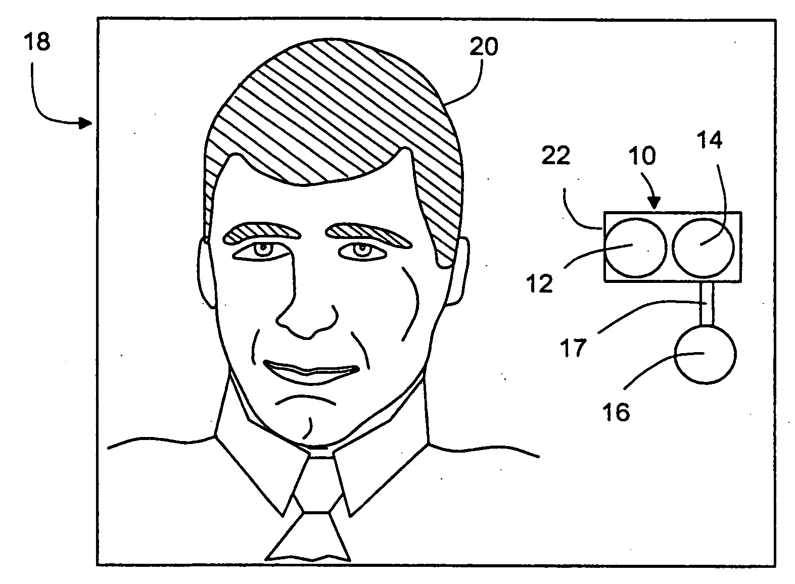
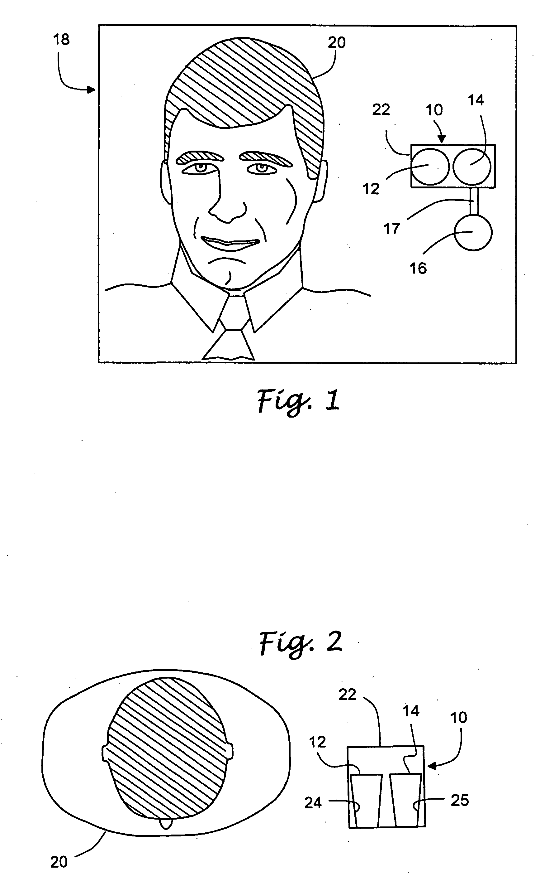
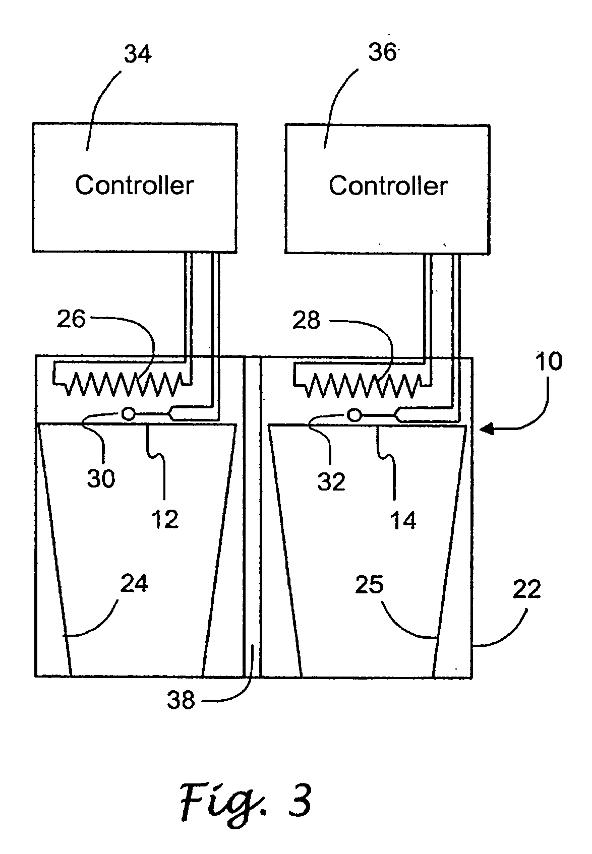
[0051]As an overview, the present invention involves acquiring thermal image data for one or more subjects and then using the thermal image data to derive temperature information about the subject(s). The acquisition of thermal image data involves using appropriate equipment and optionally the use of suitable reference temperature information to help enhance the accuracy of the acquired data. The derivation of core body temperature information may involve one or more of data calibration (such as with respect to the reference temperature data), image processing, identifying area(s) of the i...


PUM


Abstract

An approach to noninvasively, remotely and accurately detect core body temperature in a warm-blooded subject, human or animal, via thermal imaging. Preferred features such as the use of in-frame temperature references, specific anatomical target regions and a physiological heat transfer model help the present invention to overcome pitfalls inherent with existing thermal imaging techniques applied to physiological screening applications. This invention provides the ability to noninvasively, remotely and rapidly screen for diseases or conditions that are characterized by changes in core body temperature. One human application of this invention is the remote screening for severe acute respiratory syndrome (SARS), since fever is a common, early symptom. Other diseases and conditions that affect the core body temperature of humans or animals may also be noninvasively and remotely detected with this invention.

Description

PRIORITY CLAIM[0001]The present patent application is a continuation of U.S. patent application Ser. No. 10 / 854,574, filed May 25, 2004, titled METHODS AND APPARATUS FOR A REMOTE, NONINVASIVE TECHNIQUE TO DETECT CORE BODY TEMPERATURE IN A SUBJECT VIA THERMAL IMAGING, which application claims priority under 35 USC §119(e) from U.S. Provisional Patent Application having Ser. No. 60 / 471,747, filed on May 27, 2003, in the name of Gary L. McQuilkin and titled METHODS AND APPARATUS FOR A REMOTE, NONINVASIVE TECHNIQUE TO DETECT OF CORE BODY TEMPERATURE IN A SUBJECT VIA THERMAL IMAGING and bearing Attorney Docket No. 01420.0004-US-PI, wherein said patent applications are commonly owned by the assignee of the present application and wherein the entire contents of said U.S. patent applications are incorporated herein by reference.FIELD OF THE INVENTION[0002]This invention is directed to an approach to noninvasively and remotely detect core body temperature in a warm-blooded subject. More spec...

Claims


Application Information

Patent Timeline
Patent Type & Authority Applications(United States)
IPC IPC(8): A61B5/01A61B5/00
CPCA61B5/015G01J5/0025G01J2005/0077G01J5/522G01J5/53A61B5/01
Inventor MCQUILKIN, GARY L.
Owner MCQUILKIN GARY L
Why Eureka
Try Eureka