
Method and system for image segmentation using models

a model and image technology, applied in the field of medical imaging, can solve the problems of unreliable intensity based segmentation tools and difficult heart segmentation

Active Publication Date: 2008-08-07
SALIENT IMAGING +1
View PDF21 Cites 78 Cited by
  • Summary
  • Abstract
  • Description
  • Claims
  • Application Information

AI Technical Summary

Problems solved by technology

However, heart segmentation is challenging due a number of factors.
In addition, the unpredictability of patient metabolism and the use of new acquisition protocols, such as the saline flush protocol, further complicate the task, making intensity based segmentation tools unreliable.

Method used


Image

  • Method and system for image segmentation using models
  • Method and system for image segmentation using models
  • Method and system for image segmentation using models

Examples


Embodiment Construction

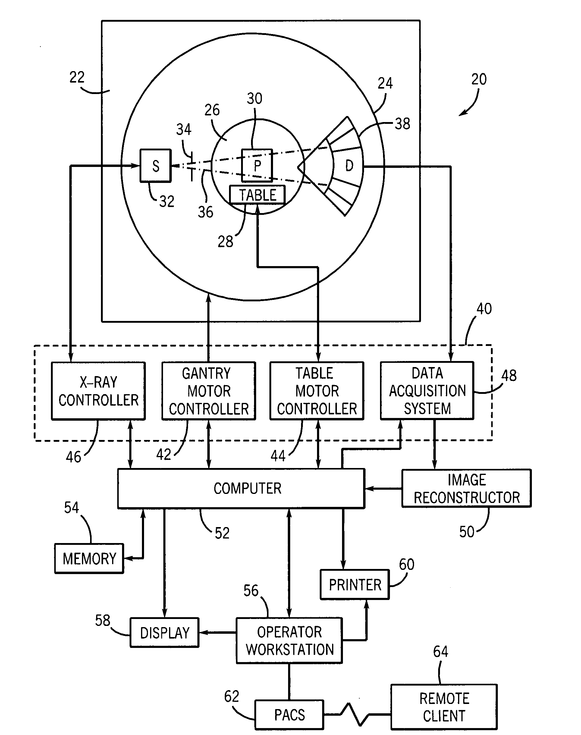
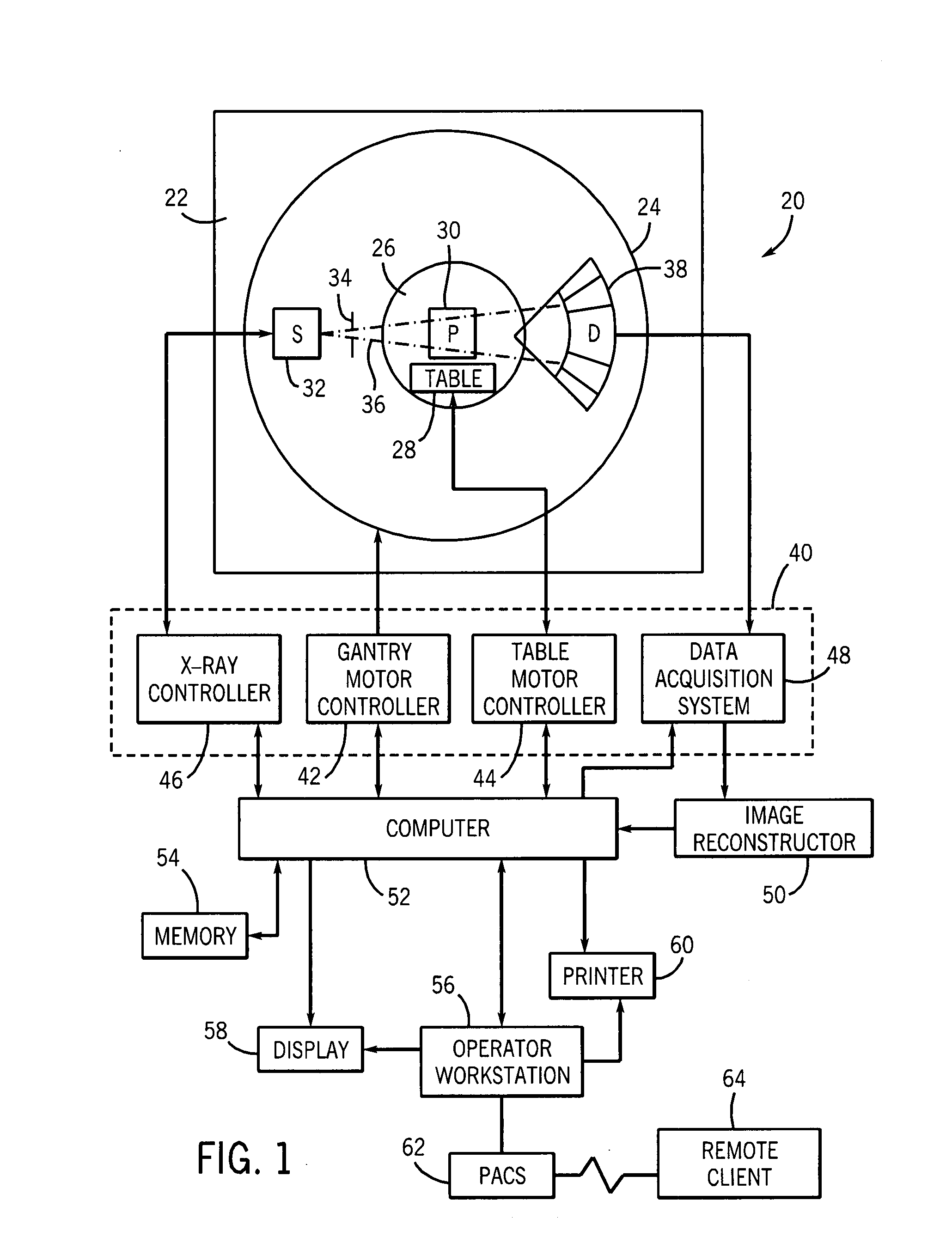
[0018]Referring now to FIG. 1, a computed tomography (CT) imaging system designed both to acquire original image data and to process the image data for display and analysis is presented, and referenced generally by reference numeral 20. The illustrated embodiment of the CT imaging system 20 has a frame 22, a gantry 24, and an aperture (imaging volume or CT bore volume) 26. A patient table 28 is positioned in the aperture 26 of the frame 22 and the gantry 24. The patient table 28 is adapted so that a patient 30 may recline comfortably during the examination process.

[0019]The illustrated embodiment of the CT imaging system 20 has an X-ray source 32 positioned adjacent to a collimator 34 that defines the size and shape of the X-ray beam 36 that emerges from the X-ray source 32. In typical operation, the X-ray source 32 projects a stream of radiation (an X-ray beam) 36 towards a detector array 38 mounted on the opposite side of the gantry 24. All or part of the X-ray beam 36 passes thro...


PUM


Abstract

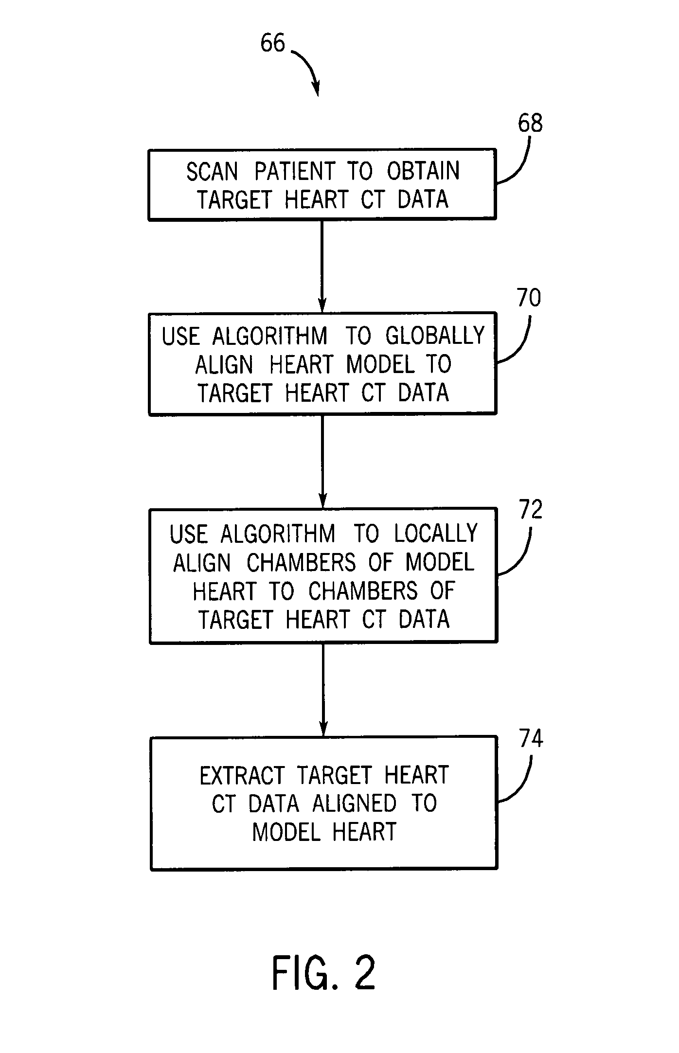
A technique is provided for utilizing a model of an anatomical feature to facilitate the segmentation of the anatomical feature from its background in a medical image. A global alignment of the model with a region in the patient's image data that generally corresponds to the anatomical feature is performed in one embodiment. Internal structural features within the model are then aligned with their corresponding structural features in the patient's image data. The portion of the patient's image data that is aligned with the model of the anatomical feature is then segmented from the remaining portions of the patient's image data that are not aligned with the model.

Description

BACKGROUND[0001]The invention relates generally to medical imaging, and more particularly to a technique for image segmentation using a model.[0002]Medical imaging has become an extremely valuable tool in the diagnosis ad treatment of many illnesses and diseases. For example, cardiac imaging using computed tomography (CT) is emerging as the protocol of choice for the diagnosis and treatment of cardiovascular disease. In addition to standard X-ray systems that produce an image on a film, medical imaging systems are now available that produce digital images that may be displayed on a monitor.[0003]Digital imaging processing enables medical images to be enhanced through the use of computers. Digital image processing has many of the same advantages in signal processing over analog image processing as does digital audio processing over analog audio processing. In addition, digital image processing enables the use of algorithms to perform other tasks, such as three-dimensional visualizati...

Claims


Application Information

Patent Timeline
Patent Type & Authority Applications(United States)
IPC IPC(8): A61B5/00
CPCA61B6/503A61B6/504G06T2207/30048G06T2207/10081G06T7/0089G06T7/149G06T7/12
Inventor SURYANARAYANAN, SRIKANTHMULLICK, RAKESHMORE, MITALI JANARDANSHRIRAM, KRISHNA SEETHARAM
Owner SALIENT IMAGING
Learn More