Ophtalmologic surgical microscope having a measuring unit

a surgical microscope and measuring unit technology, applied in the field of surgical microscopes for ophthalmology, can solve the problems of inability to reliably determine the optical state of the eye, inability to achieve the optical effect, and possible partial amplification but also mutual weakening of the optical effect, so as to achieve substantial advantages for patients

Active Publication Date: 2008-08-21
CARL ZEISS SURGICAL
View PDF11 Cites 24 Cited by
  • Summary
  • Abstract
  • Description
  • Claims
  • Application Information

AI Technical Summary

Benefits of technology

[0025]The surgical microscope of the invention makes possible a display of a model and corresponding characteristic variables during the course of a surgical procedure. The measurement data for the model are determined during the course of surgery in order to be able to recognize an unwanted deviation from a planned state of the patient eye and to provide the surgeon with the capability to correspondingly change the surgical procedure as may be required thereby affording very substantial advantages for the patient.

Problems solved by technology

These visual deficiencies are caused by the geometric optical correction of the eye which is by nature inadequate.
However, the optical state of the eye cannot be reliably determined on the basis of this single optical characteristic variable.
It has been shown that, in the course of a healing process after a surgical procedure on the eye, not only a single optically effective variable changes but a modification of several optical characteristic variables of the eye takes place whose optical effect possibly partially amplifies but also mutually weakens.
On the other hand, this is caused by the situation that the optical components of the eye, namely cornea and lens, are far from perfect.
This leads to the situation that the object is observed by the eye as being blurred.
This elasticity has the consequence that, with the human eye, it is not at all possible to image a point precisely as a point.
This is problematic because, during the course of surgery, and during a healing of the wound, optical characteristic variables of the eye can change such as distances of refractive elements.

Method used


Image

  • Ophtalmologic surgical microscope having a measuring unit
  • Ophtalmologic surgical microscope having a measuring unit
  • Ophtalmologic surgical microscope having a measuring unit

Examples


Embodiment Construction

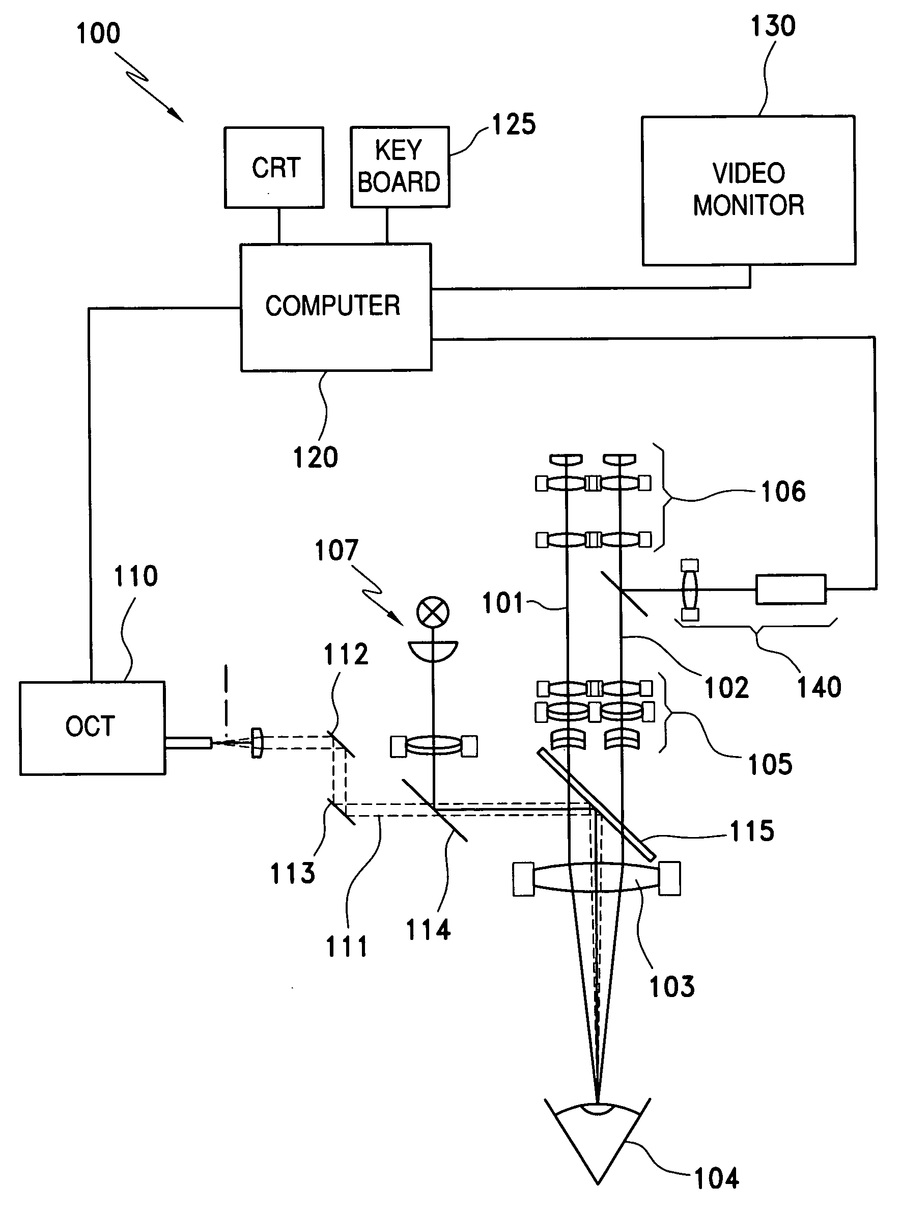
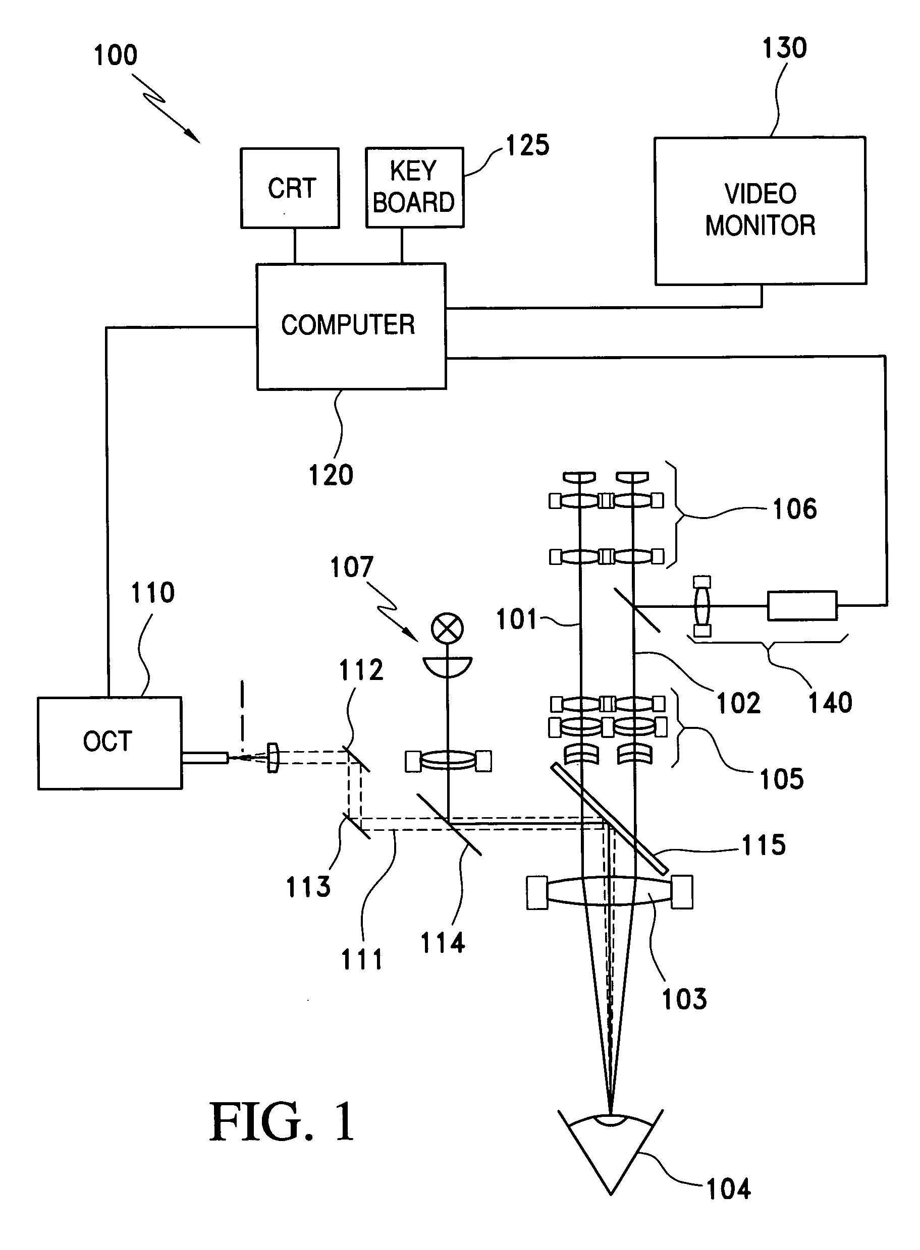
[0030]The ophthalmologic surgical microscope 100 of FIG. 1 includes a stereoscopic illuminating beam path (101, 102) which makes possible the examination of a patient eye 104 through a microscope main objective 103. The ophthalmologic surgical microscope 100 further has a zoom system 105 and an ocular 106. The surgical microscope 100 includes an illuminating system 107 which makes available illuminating light for the patient eye 104 through the microscope main objective 103.

[0031]As a measuring device for determining optical characteristic variables of the patient eye 104, the ophthalmologic surgical microscope 100 has a coherence interferometer in the form of an OCT-system 110. The OCT-system 110 provides a scanning light beam 111 of short coherent light which is guided to the patient eye 104 via displaceable scan mirrors (112, 113) and beam splitters (114, 115) through the microscope main objective 103. The light of the scanning light beam 111, which is scattered at the patient ey...


PUM


Abstract

A surgical microscope (100) for ophthalmology includes a measuring unit (110) for determining at least one characteristic value of a patient eye (104). The measuring unit (110) is connected to a computer unit (120) which calculates a model of the patient eye (104) based on the determined characteristic variables. The surgical microscope also includes a display device for displaying the calculated model of the patient eye (104) or characteristic variables that are derived from the calculated model.

Description

CROSS REFERENCE TO RELATED APPLICATIONS[0001]This application is a continuation-in-part application of international patent application PCT / EP 2006 / 008616, filed Sep. 5, 2006, designating the United States and claiming priority from German application 10 2005 042 436.8, filed Sep. 7, 2005, and the entire content of both applications is incorporated herein by reference.FIELD OF THE INVENTION[0002]The invention relates to a surgical microscope for ophthalmology having a measuring unit for determining at least one optical characteristic variable of the eye of a patient.BACKGROUND OF THE INVENTION[0003]The geometric length between optically effective surfaces of the eye is understood to be included in the optical characteristic variables of an eye of a patient whereat a jump or a non-vanishing gradient of the index of refraction occurs or whereat an absorption of light takes place. The local radii of the surfaces, that is, their Gaussian curvatures can, however, likewise be considered a...

Claims


Application Information

Patent Timeline
Patent Type & Authority Applications(United States)
IPC IPC(8): A61B3/10
CPCA61B3/0025A61B3/13A61B19/52G02B21/36A61B19/5225G02B21/0012A61B19/5223A61B90/36A61B90/20A61B90/37
Inventor GAIDA, GERHARD
Owner CARL ZEISS SURGICAL
Why Eureka
Try Eureka