
Fluid-fillable ultrasound imaging catheter tips

Inactive Publication Date: 2008-11-20
GENERAL ELECTRIC CO
View PDF31 Cites 15 Cited by
  • Summary
  • Abstract
  • Description
  • Claims
  • Application Information

AI Technical Summary

Benefits of technology

[0006]Briefly, in accordance with aspects of the present technique, an imaging catheter tip is presented. The imaging catheter tip includes a housing. Further, the imaging catheter tip includes a transducer assembly located within the housing in a distal portion of the imaging catheter tip. The transducer assembly includes a transducer. In addition, the imaging catheter tip includes a motor located within the housing in a proximal portion of the imaging catheter tip. The motor is configured to facilitate oscillation of the transducer assembly about a longitudinal axis of the imaging catheter tip. The imaging catheter tip also includes a fill tube disposed between the motor and the housing. The fill tube is configured to deliver acoustic coupling fluid to the distal portion of the imaging ca

Problems solved by technology

Such applications are quite demanding, requiring very small transducer packages that can nevertheless collect large amounts of information.
However, it may be difficult to provide a transducer package that is mechanically, acoustically, and electrically suitable.

Method used


Image

  • Fluid-fillable ultrasound imaging catheter tips
  • Fluid-fillable ultrasound imaging catheter tips
  • Fluid-fillable ultrasound imaging catheter tips

Examples


Embodiment Construction

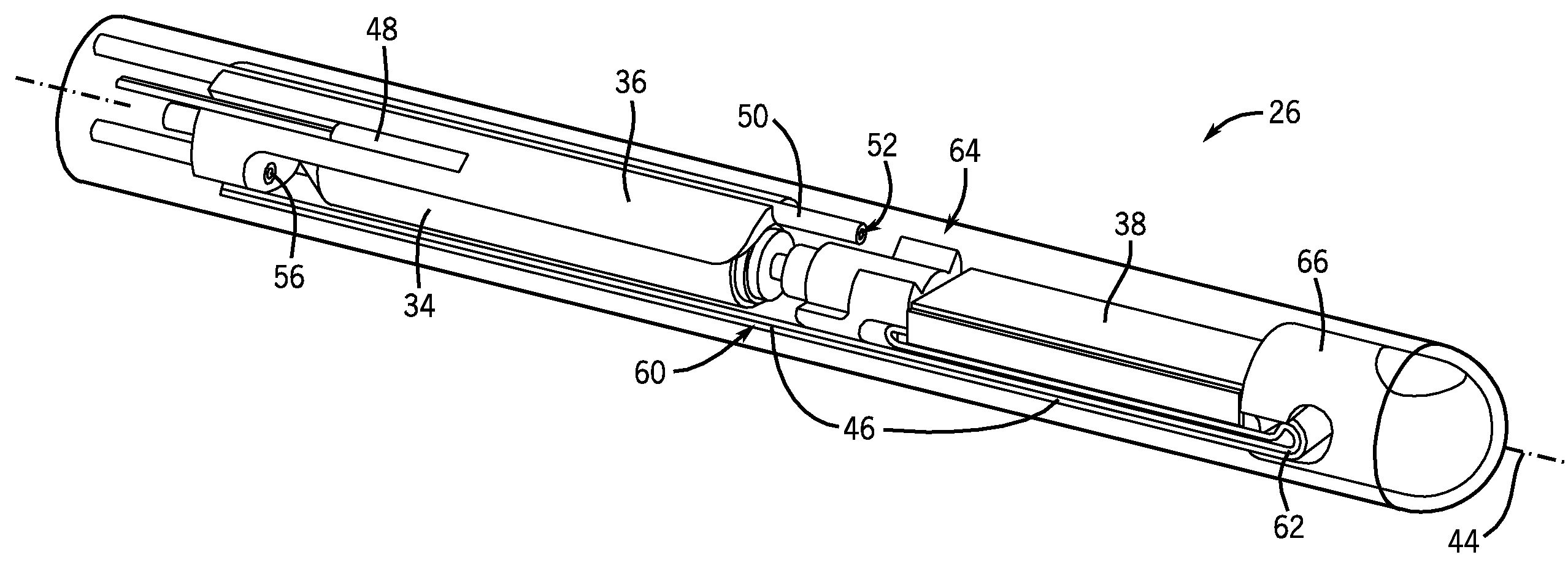
[0017]Imaging probes containing transducer assemblies for obtaining real-time, three-dimensional ultrasound imaging may use a motor assembly to oscillate transducer assemblies about an axis of the imaging probe. In doing so, the transducer assemblies may be capable of obtaining real-time, three-dimensional ultrasound images by sweeping a two-dimensional slice through a three-dimensional volume. Acoustic coupling fluid may be used in conjunction with the transducer assemblies to provide an effective or suitable acoustic transition between the transducer and the surrounding housing. Such imaging probes are typically filled with acoustic coupling fluid at the time of manufacture. Such imaging probes also typically include a fluid barrier to separate the motor from the fluid-filled space that houses the transducer. The barrier is penetrated by a driveshaft that couples the motor to the moving transducer. A fluid seal on the driveshaft prevents or minimizes the leakage of fluid from the ...


PUM


Abstract

An imaging catheter tip is presented. The imaging catheter tip includes a housing. Further, the imaging catheter tip includes a transducer assembly located within the housing in a distal portion of the imaging catheter tip. The transducer assembly includes a transducer. In addition, the imaging catheter tip includes a motor located within the housing in a proximal portion of the imaging catheter tip. The motor is configured to facilitate oscillation of the transducer assembly about a longitudinal axis of the imaging catheter tip. The imaging catheter tip also includes a fill tube disposed between the motor and the housing. The fill tube is configured to deliver acoustic coupling fluid to the distal portion of the imaging catheter tip.

Description

CROSS REFERENCE TO RELATED APPLICATIONS[0001]This application claims priority to U.S. Provisional Patent Application No. 60 / 917,995, entitled “SELF-CONTAINED, FLUID-FILLABLE REAL-TIME THREE-DIMENSIONAL ULTRASOUND IMAGING CATHETER TIPS,” filed May 15, 2007, which is herein incorporated by reference in its entirety for all purposes.BACKGROUND[0002]The present disclosure relates generally to ultrasound imaging and, more particularly, to imaging catheter tips which contain acoustic transducers for obtaining ultrasound images.[0003]Acoustic transducers have found application in medical imaging where an acoustic probe is held against a patient and the probe transmits and receives ultrasound waves. The received energy may, in turn, facilitate the imaging of the tissues of the patient. For example, transducers may be employed to image the heart of the patient.[0004]Catheter-based ultrasonic imaging techniques are interventional procedures that generally involve inserting a probe, such as an...

Claims


Application Information

Patent Timeline
IPC IPC(8): A61B8/14A61B8/12
CPCA61B8/12A61B8/4281A61B8/445A61B8/4461A61B8/483
Inventor LEE, WARRENWILDES, DOUGLAS GLENNGRIFFIN, WESTON BLAINEBRUESTLE, REINHOLD
Owner GENERAL ELECTRIC CO
Features
  • Generate Ideas
  • Intellectual Property
  • Life Sciences
  • Materials
  • Tech Scout
  • Unparalleled Data Quality
  • Higher Quality Content
  • 60% Fewer Hallucinations
Learn More