Method and Apparatus for Volume Rendering of Medical Data Sets

a technology for medical data and volume rendering, applied in the field of method and apparatus for volume rendering of medical data sets, can solve the problems of time-consuming and complicated adjustment of viewport parameters, and achieve the effect of reducing the difficulty of adjusting the viewport parameters and reducing the difficulty of adjusting the parameters

Inactive Publication Date: 2009-01-01
AGFA HEALTHCARE NV
View PDF5 Cites 45 Cited by
  • Summary
  • Abstract
  • Description
  • Claims
  • Application Information

AI Technical Summary

Benefits of technology

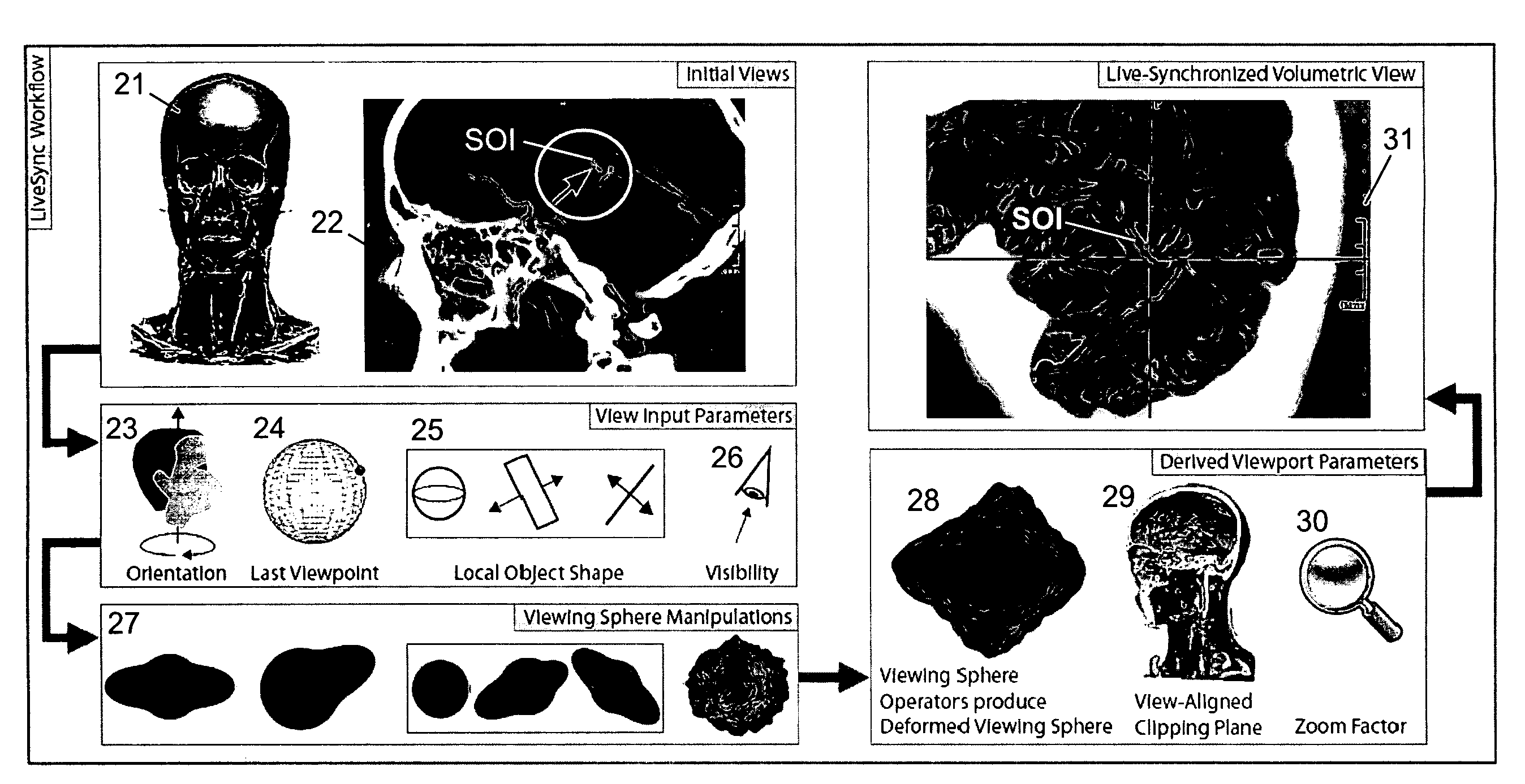
[0055]It is preferred that said at least two deformed viewing spheres obtained from the previously acquired medical data sets are superimposed so that an accumulated deformed viewing sphere is obtained and that the first parameters are derived from said accumulated deformed viewing sphere. In this preferred embodiment, the information contained in different deformed viewing spheres calculated from medical data of the same or different patients but originating from the same medical examination type, e.g. computed tomography images of patients' heads, is accumulated or superimposed, e.g. by adding and / or averaging, in the accumulated deformed viewing sphere. From said accumulated deformed viewing sphere first parameters resulting in a good initial view of the currently acquired medical data of the currently examined patient can be deduced easily and very quickly.

Problems solved by technology

In spite of the research it is still complicated and time-consuming to adjust the viewport parameters in order to achieve a good and unobstructed view on the structure of interest.

Method used


Image

  • Method and Apparatus for Volume Rendering of Medical Data Sets
  • Method and Apparatus for Volume Rendering of Medical Data Sets
  • Method and Apparatus for Volume Rendering of Medical Data Sets

Examples


application examples

[0123]Preferably, the invention is executed on a medical computer workstation. The corresponding computations for the LiveSync viewpoint selection can be performed interactively and are not influenced very much by the size of the medical data set. E.g., on a PC configured with an AMD Athlon 64 Dual Core Processor 4400+ and 2 GB of main memory the LiveSync-related computations take about 70 ms to 150 ms per picking, depending on the number of segmented voxels at the local segmentation step and on the estimated local feature shape. Thus, the user gets an almost instant update of the volumetric view whenever he or she is picking on a certain structure in a 2D slice. To demonstrate the usefulness of the interactively synchronized views the results for three different application scenarios will be discussed in the following.

[0124]In a first example an estimated good viewpoint and a rather bad viewpoint are presented for comparison reasons. FIG. 8 shows the results for a picking action on...


PUM


Abstract

A method and a corresponding apparatus for volume rendering of medical data sets comprises a display (11) for displaying at least one slice view (12) of a medical data set (18) of an object, in particular a patient. A user selection unit (13, 14, 15) enables a user to select a position on a structure of interest (SOI) in the at least one displayed slice view (12), a volume rendering unit (16) for determines a volume rendering (17) of the structure of interest (SOI) based on one or more first parameters. The first parameters characterize the view of the displayed volume rendering (17, 31) of the structure of interest (SOI), the volume rendering (17, 31) of the structure of interest (SOI) being displayed on the display (11). In order to achieve good volumetric views of the medical data set (18) requiring reduced user input the volume rendering unit (16) is designed for determining the first parameters automatically by considering the user-selected position and one or more second parameters, the second parameters characterizing at least one of the object, the structure of interest (SOI), the currently displayed slice view (12) and one or more previous volume renderings (17) of the structure of interest (SOI), and the volume rendering unit (16) is designed for determining the first parameters in that way that an optimized view on the displayed volume rendering (17) of the structure of interest (SOI) is achieved without additional user input apart from the user-selection of the position.

Description

RELATED APPLICATIONS[0001]This application claims priority to European Patent Application No EP07118075.6, filed on Oct. 9, 2007, which is incorporated herein by reference in its entirety.BACKGROUND OF THE INVENTION[0002]Modern modalities for medical imaging (e.g. computed tomography) provide large quantities of data at an unprecedented resolution. Presenting this enormous amount of information is a challenging task for today's radiology workstations. Volumetric rendering is the current method of choice for providing a good survey of the data. Combining the information provided by two-dimensional (2D) cross-sections and three-dimensional (3D) visualization can improve the diagnosis process. Linking the different representations of the data has the potential benefit to provide significant enhancements in efficiency. Usually the volumetric display acts as an overview display in this context. The cross-sectional images contain diagnostically relevant information.[0003]Pinpointing a pat...

Claims


Application Information

Patent Timeline
Patent Type & Authority Applications(United States)
IPC IPC(8): G06T15/00G06T15/08
CPCA61B6/032G06T15/08A61B6/466A61B6/463
Inventor KANITSAR, ARMINKOHLMANN, PETERBRUCKNER, STEFANGROELLER, M. EDUARDWEGENKITTL, RAINERMROZ, LUKAS
Owner AGFA HEALTHCARE NV
Why Eureka
Try Eureka