Mapping of Retinal Parameters from Combined Fundus Image and Three-Dimensional Optical Coherence Tomography

a three-dimensional optical coherence tomography and combined fundus image technology, applied in the field of ophthalmic characterization, can solve the problems of difficult static process, difficult to relate numerical retinal thickness values, tabular or grid format,

Active Publication Date: 2009-05-14
TOPCON MEDICAL SYST
View PDF15 Cites 48 Cited by
  • Summary
  • Abstract
  • Description
  • Claims
  • Application Information

AI Technical Summary

Problems solved by technology

Correlating numerical values of retinal thickness, either in tabular or grid format, with anatomical features is a difficult and, at best, a static process.

Method used


Image

  • Mapping of Retinal Parameters from Combined Fundus Image and Three-Dimensional Optical Coherence Tomography
  • Mapping of Retinal Parameters from Combined Fundus Image and Three-Dimensional Optical Coherence Tomography
  • Mapping of Retinal Parameters from Combined Fundus Image and Three-Dimensional Optical Coherence Tomography

Examples


Embodiment Construction

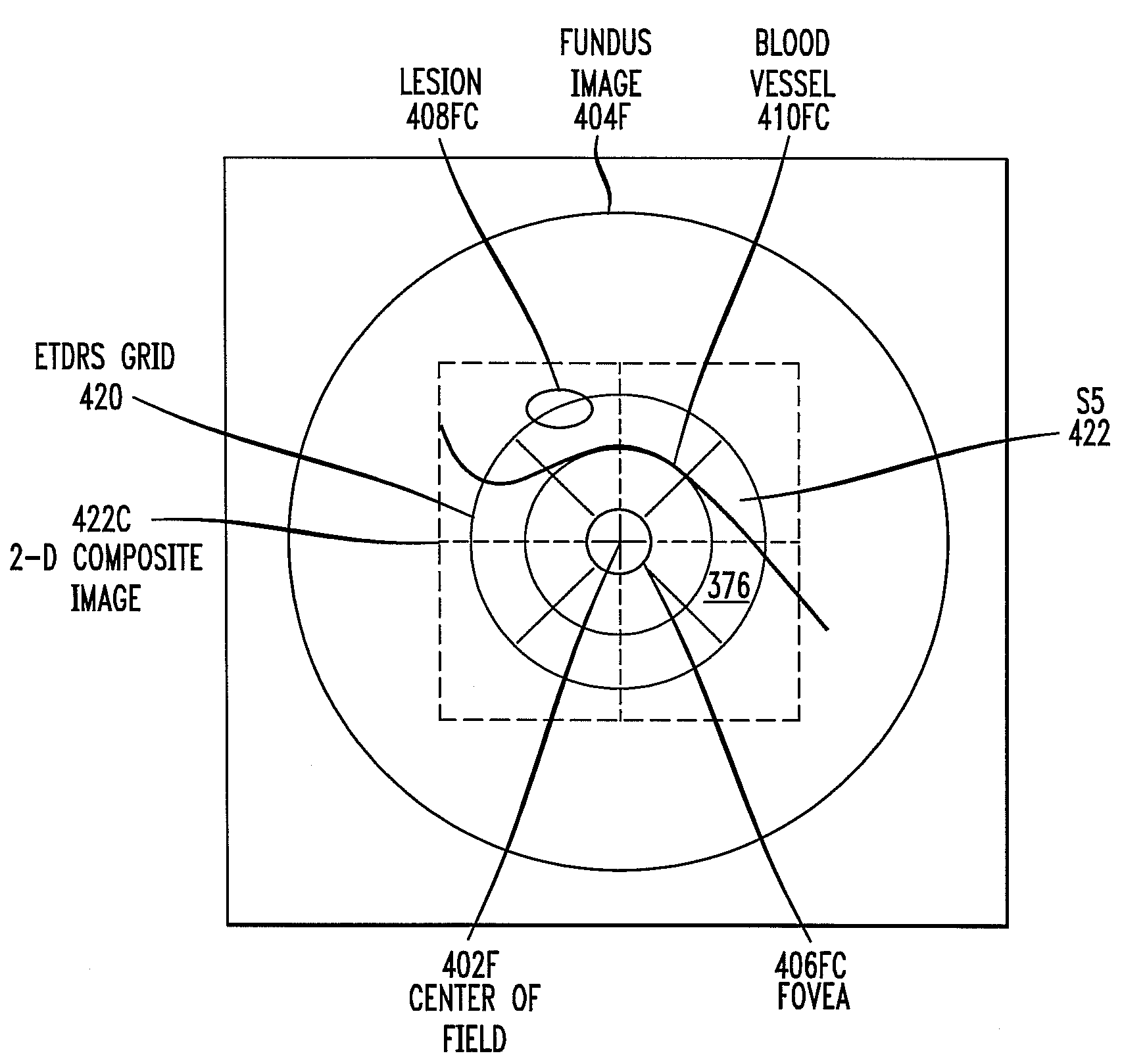
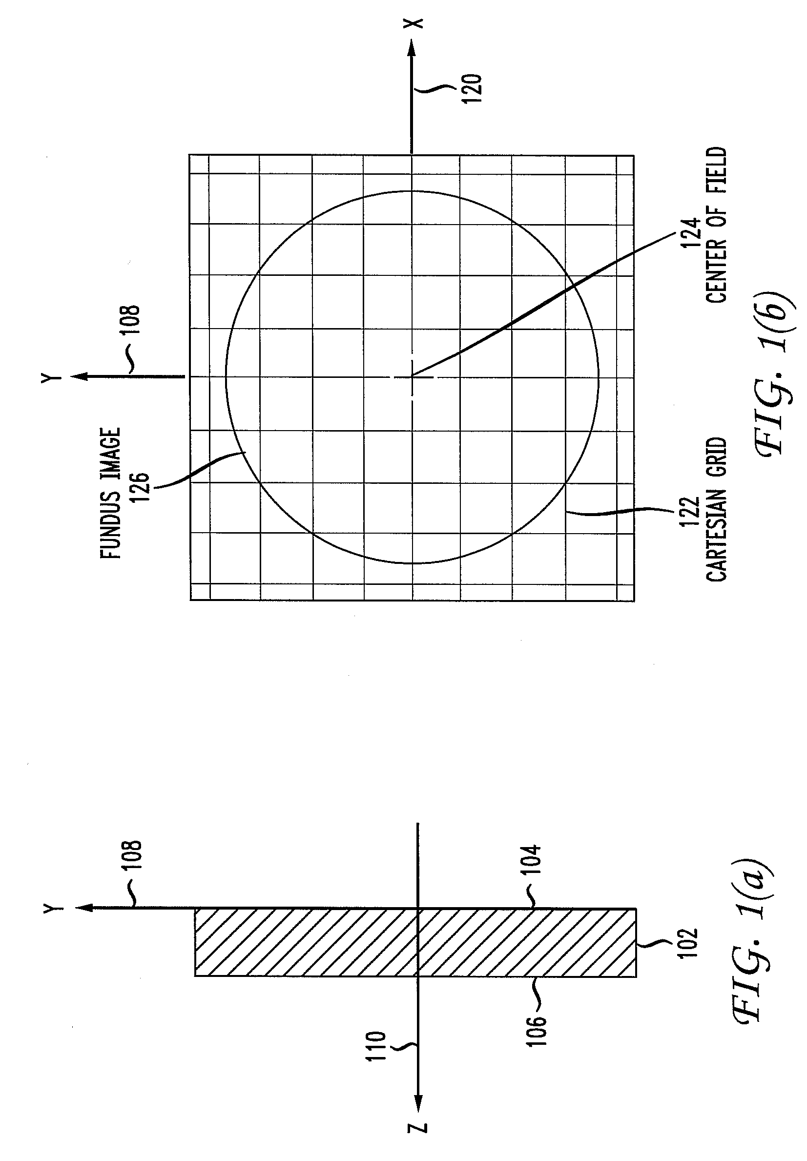
[0022]Diagnostics for eye disorders typically include a detailed ophthalmic examination of the retina. For initial examination, an eye doctor will view the retina through an ophthalmoscope. For a permanent record, the retina is typically photographed with a fundus camera. A fundus photograph directly records various anatomical features of the retina, such as the optic disc, fovea, blood vessels, and lesions. The image may be captured on film or stored in digital form and displayed on a monitor. Visual inspection of the retina continues to be a primary diagnostic technique.

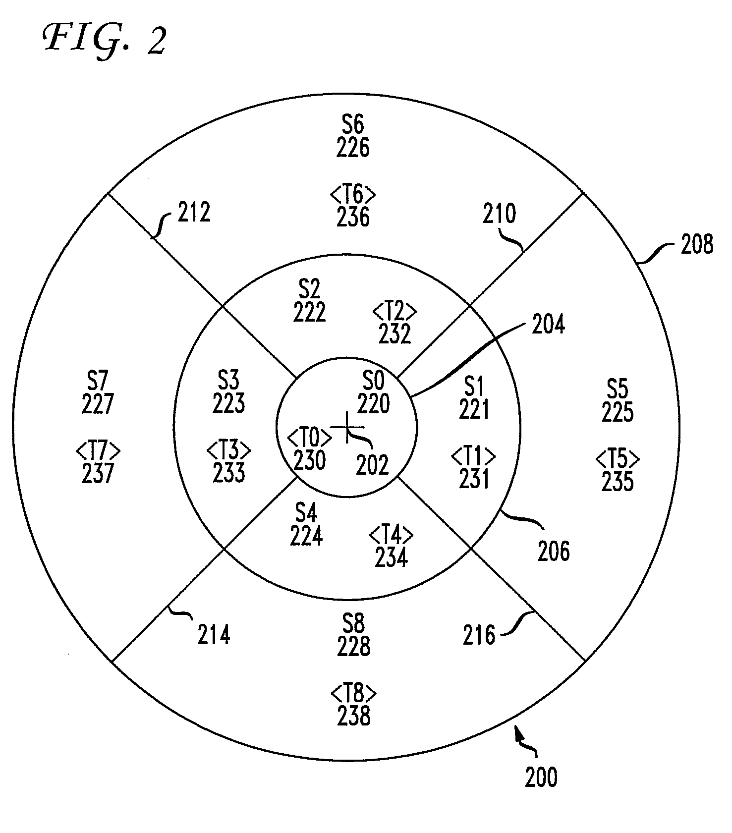
[0023]More sophisticated techniques have recently been developed for diagnostics of the eye. A powerful technique for characterizing and imaging ocular structures, including the retina, is three-dimensional optical coherence tomography (3-D OCT). In this technique, an optical probe, typically a laser beam, is directed onto the retina. Part of the beam is back-reflected. Interferometric analysis of the back-reflecte...


PUM


Abstract

A second retinal characterization data set is mapped to a first retinal characterization dataset. The first retinal characterization dataset is displayed as a first graphical map. The second retinal characterization dataset is displayed as a second graphical map which is mapped to the first graphical map. The second graphical map may be warped and morphed onto the first graphical map. Retinal characterization datasets may be derived either from a fundus image or from a retinal parameter dataset calculated from a three-dimensional optical coherence tomography scan of a retina. Retinal parameter datasets may characterize parameters such as retinal thickness. In an embodiment, a fundus image is warped and morphed onto a retinal surface topographical map.

Description

[0001]This application claims the benefit of U.S. Provisional Application No. 61 / 002,363 filed Nov. 8, 2007, which is incorporated herein by reference.BACKGROUND OF THE INVENTION[0002]The present invention relates generally to ophthalmic characterization, and more particularly to mapping retinal parameters from combined fundus image and three-dimensional optical coherence tomography.[0003]Diagnostics for eye disorders typically include a detailed ophthalmic examination of the retina. For initial examination, an eye doctor will view the retina through an ophthalmoscope. For a permanent record, the retina is typically photographed with a fundus camera. A fundus photograph directly records various anatomical features of the retina, such as the optic disc, fovea, blood vessels, and lesions. The imaging capabilities of fundus photography may be enhanced by supplementary techniques. A high-contrast image of retinal blood vessels, for example, may be photographed after the injection of a f...

Claims


Application Information

Patent Timeline
Patent Type & Authority Applications(United States)
IPC IPC(8): G06K9/00
CPCA61B3/0058A61B3/1005A61B5/7425A61B3/145A61B3/1225
Inventor HUANG, YIJUNROYAMA, TETSUYOSHIKOTCHKIN, ALEXANDRE
Owner TOPCON MEDICAL SYST
Why Eureka
Try Eureka