
Compositions and methods for detection of antibody binding to cells

a technology of antibody binding and cell, applied in the field of binding proteins, can solve the problems of human antibody in vitro, ebv)-mediated transformation or cell fusion, and many proteins cannot be purified in a non-denatured sta

Inactive Publication Date: 2009-05-14
THE TRUSTEES OF THE UNIV OF PENNSYLVANIA
View PDF5 Cites 24 Cited by
  • Summary
  • Abstract
  • Description
  • Claims
  • Application Information

AI Technical Summary

Problems solved by technology

In addition, there exists certain situations where, for diagnostic purposes, it is essential that human antibodies be used because other animals are unable to make antibodies against the antigen to be detected in the diagnostic method.
For example, in order to determine the Rh phenotype of human red blood cells (RBCs), human sera that contains anti-Rh antibody must be used since other animals cannot make an antibody capable of detecting the human Rh antigen.
The production of human antibodies in vitro by immortalizing human B lymphocytes using Epstein Barr virus (EBV)-mediated transformation or cell fusion has been fraught with technical difficulties due to the relatively low efficiency of both EBV-induced transformation and cell fusion when compared with the murine system.
There are several difficulties associated with the generation of antibodies using bacteriophage.
For example, many proteins cannot be purified in a non-denatured state, in that purification procedures necessarily involve solubilization of protein which may render some proteins permanently denatured with concomitant destruction of antigenic sites present thereon.
Such proteins thus cannot be bound to a solid phase and therefore cannot be used to pan for phage bearing antibodies which bind to them.
However, it was discovered that since phage are inherently “sticky” and RBCs express a multitude of antigens on the cell surface, a sufficient amount of phage which do not express the appropriate antibody on the surface also adhere to the RBCs, thus rendering the method impractical for isolation of phage which express antibody of desired specificity.
The isolation of fluorescently labeled cells is accomplished using the technique of fluorescently-activated cell sorting (FACS), an expensive and time-consuming procedure.

Method used


Image

  • Compositions and methods for detection of antibody binding to cells
  • Compositions and methods for detection of antibody binding to cells
  • Compositions and methods for detection of antibody binding to cells

Examples


example 1

Isolation of Cell Surface-Specific Human Monoclonal Antibodies Using Phage Display and Magnetically-Activated Cell Sorting

[0120] The experiments described in this Example provide procedures and results for the isolation and production of anti-Rh(D) red blood cell antibodies using Fab / phage display.

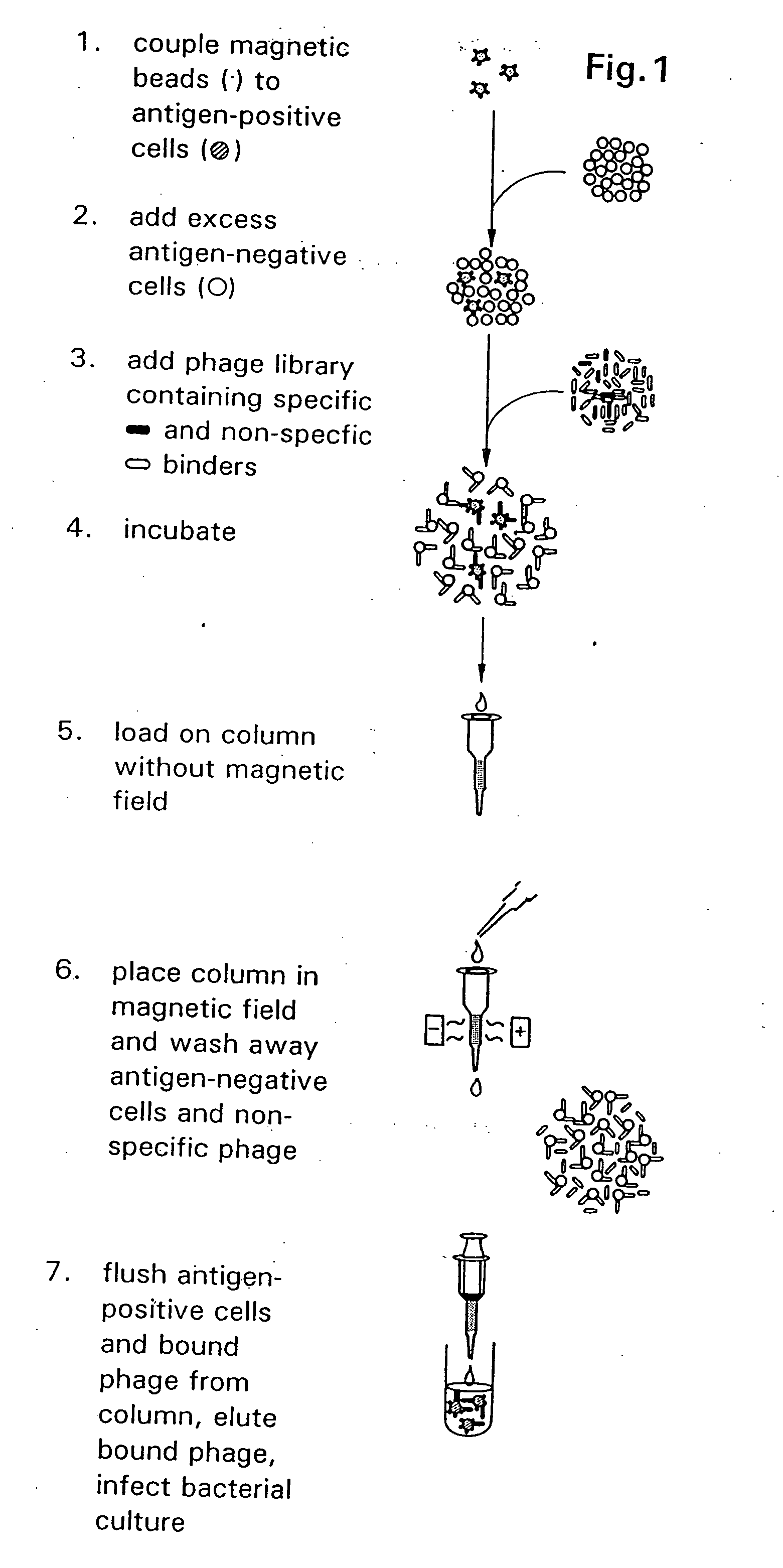
[0121] A method is described in FIG. 1 for the isolation of filamentous phage-displayed human monoclonal antibodies specific for non-purifiable cell surface expressed molecules. To optimize the capture of antigen-specific phage and minimize the binding of irrelevant phage antibodies, a simultaneous positive and negative selection strategy was employed. Cells bearing the antigen of interest are pre-coated with magnetic beads and are diluted into an excess of unmodified antigen-negative cells. Following incubation of the cell admixture with a Fab / phage library, the antigen positive cell population is retrieved using magnetically-activated cell sorting, and antigen-specific Fab / phage are el...

example 2

Genetic and Immunological Properties of Phage-Displayed Human Anti-Rh(D) Antibodies

[0172] Clinically, the human Rh(D) antigen is the most important red blood cell (RBC) membrane protein in transfusion medicine. The autoimmune response against Rh(O) produces high affinity IgG antibodies which cause hemolytic transfusion reactions and hemolytic disease of the newborn (HDN). The prophylactic use of Rh(D)-immune globulin in pregnant Rh(D)-negative women has been a major advance in the prevention of HDN, yet the mechanism by which the drug exerts its immune modulatory effect is not well understood.

[0173] Monoclonal antibodies derived from the B cells of Rh(D)-immune globulin donors have defined several dozen Rh(D) epitopes (Scott, 1996, Transfus. Clin. Biol. 3:333). Paradoxically, the Rh(D) antigen, a circa 30 kD transmembrane. protein, has minimal extracellular mass and presents a very limited surface area for epitope expression. Because molecular cloning of a large repertoire of anti...

example 3

Isolation of Anti-Rh(D) Monoclonal Antibodies to Conventional and Novel Epitopes Using a Heavy Chain / Light Chain Shuffling Approach

[0357] In view of the results obtained in Examples 1 and 2 herein, heavy and light chains of antibodies of various Rh(D) epitope specificities were randomly recombined in order to generate anti-Rh(D) antibodies having additional patterns of reactivity with Rh(D) variant cells. Using this approach, plasmid DNA obtained from the Fab / phage display libraries described in panning rounds 2 and 3 of Example 1 was randomly recombined to generate a “shuffled” Fab / phage display library. When the Rh(D) specificity of antibodies of this “shuffled” library was determine, it was found that many of these antibodies exhibited novel epitope specificity. Significantly, antibody clones having novel Rh(D) epitope specificity were identified, including clones which bind to wild type and certain partial D type red blood cells but which do not bind to D category III red blood...


PUM

PropertyMeasurementUnit
mole fractionaaaaaaaaaa

Abstract

The invention includes Rh(D) binding proteins, including antibodies, and DNA encoding such proteins. Methods of generating such proteins and DNAs are also included.

Description

CROSS REFERENCE TO RELATED APPLICATIONS [0001] This application is entitled to priority pursuant to 35 U.S.C. §119(e) to U.S. provisional patent application 60 / 081,380, which was filed on Apr. 10, 1998, and is a continuation-in-part of U.S. application Ser. No. 08 / 884,045, filed Jun. 27, 1997, which application is entitled to priority under 35 U.S.C. §119(e) to U.S. Provisional Application No. 60 / 028,550, filed on Oct. 11, 1996.GOVERNMENT SUPPORT [0002] This invention was supported in part by a grant from the U.S. Government (NIH Grant No. P50-HL54516) and the U.S. Government may therefore have certain rights in the invention.FIELD OF THE INVENTION [0003] The field of the invention is generation of binding proteins. BACKGROUND OF THE INVENTION [0004] The ability to produce monoclonal antibodies has revolutionized diagnostic and therapeutic medicine. Monoclonal antibodies are typically produced by immortalization of antibody-producing mouse lymphocytes thus ensuring an endless supply...

Claims


Application Information

Patent Timeline
Patent Type & Authority Applications(United States)
IPC IPC(8): A61K39/395C07H21/04C12P21/06C12N5/06C07K16/28C07K16/08C07K16/34C12N15/10C40B40/02
CPCC07K16/005C07K16/08C07K16/34C40B40/02C12N15/1013C12N15/1037C07K2317/55
Inventor SIEGEL, DONALD L.
Owner THE TRUSTEES OF THE UNIV OF PENNSYLVANIA
Learn More