Use of Mutant Herpes Simplex Virus-2 for Cancer Therapy
a technology of herpes simplex virus and cancer therapy, which is applied in the field of cancer biology, cell biology, molecular biology, etc., can solve the problems of increasing the destructive power of the virus against tumor cells, severe compromise of the ability of the virus to replicate in cells, etc., and achieves a strong anti-tumor immune response
- Summary
- Abstract
- Description
- Claims
- Application Information
AI Technical Summary
Benefits of technology
Problems solved by technology
Method used
Image
Examples
example 1
Construction of FusOn-H2
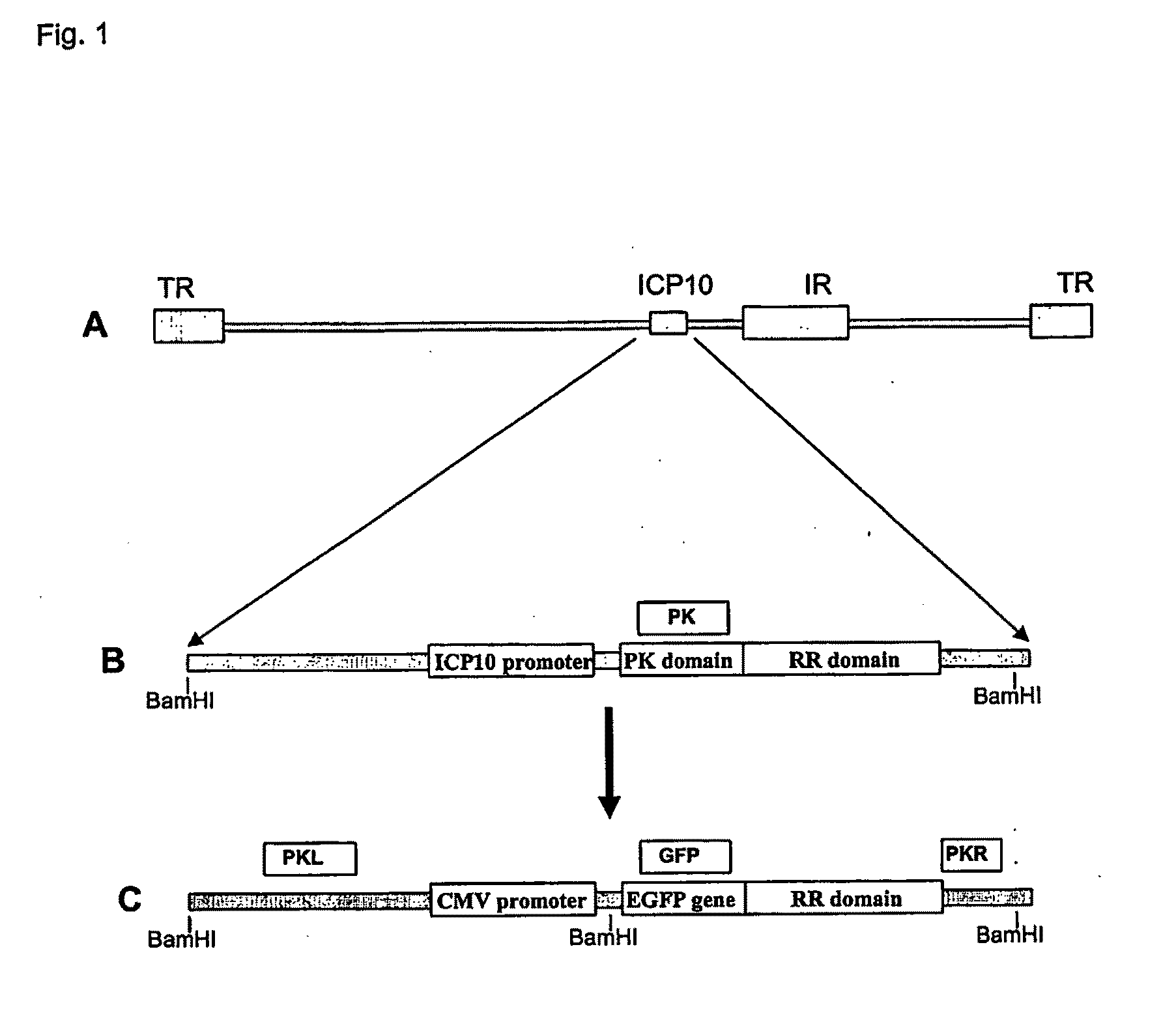
[0168]The construction of the exemplary FusOn-H2 is illustrated in FIG. 1. Initially the HSV genome region comprising the ICP10 left-flanking region (equivalent to nucleotide number of HSV-2 genome 85994-86999) was amplified with the following exemplary pair of primers: 5′-TTGGTCTTCACCTACCGACA (SEQ ID NO:1); and 3′-GACGCGATGAACGGAAAC (SEQ ID NO:2). The RR domain and the right-flank region (equivalent to the nucleotide sequence number of HSV-2 genome 88228-89347) were amplified with the following exemplary pair of primers: 5′-ACACGCCCTATCATCTGAGG (SEQ ID NO:13); and 5′-AACATGATGAAGGGGCTTCC (SEQ ID NO:14). These two PCR products were cloned into pNeb 193 through EcoRI-NotI-XbaI ligation to generate pNeb-ICP10-deltaPK. Then, the DNA sequence containing the CMV promoter-EGFP gene was PCR amplified from pSZ-EGFP with the following exemplary pair of primers: 5′-ATGGTGAGCAAGGGCGAG (SEQ ID NO:3); and 3′-CTTGTACAGCTCGTCCATGC (SEQ ID NO:4). The PCR-amplified DNA was th...
example 2
In Vitro Characterization
[0170]The exemplary FusOn-H2 vector was characterized by standard methods in the art.
Southern Blot Analysis.
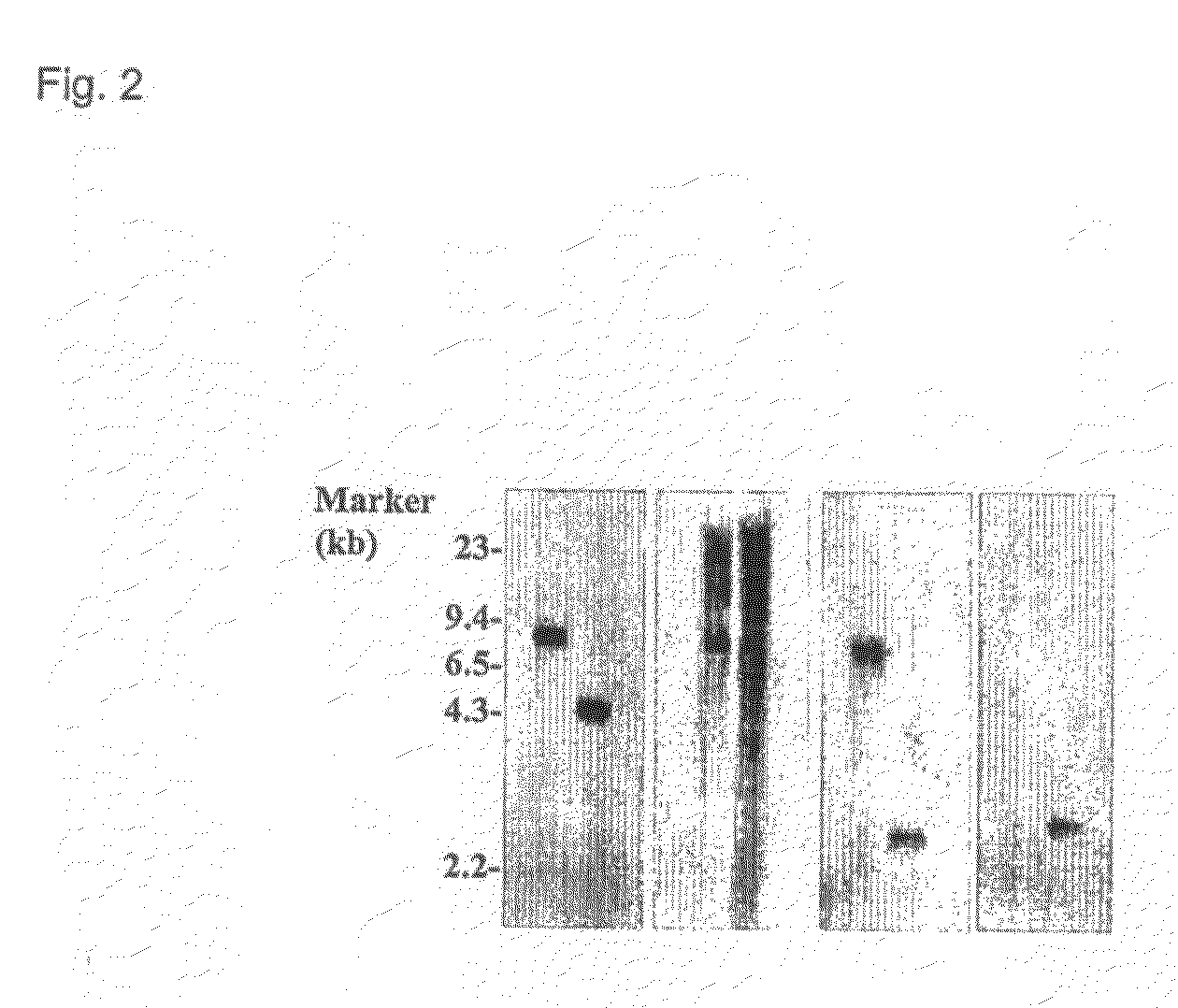
[0171]To confirm that the modified ICP10 gene has been correctly inserted into the HSV-2 genome to replace the original ICP10 gene, virion DNA was extracted from purified FusOn-H2 virus stock. As a control, virion DNA from the parental wild type HSV-2 was extracted according to the same procedure. The virion DNA was digested with BamHI and electrophoresed in an 0.8% agarose gel. BamHI digestion generates an 7390 bp DNA fragment from the wild type HSV-2 genome that comprises the entire ICP10 gene and its left and right flank regions. However, digestion of FusOn-H2 genome by the same enzyme generates two DNA fragments from the ICP10 gene locus: 1) a 4830 bp fragment comprising the left-flank and the CMV promoter sequence; and 2) a 3034 bp sequence comprising the GFP, RR, and the right-flank region. The DNA was transferred to a nylon membrane and hybridiz...
example 3
In Vitro Phenotypic Characterization of FusOn-H2
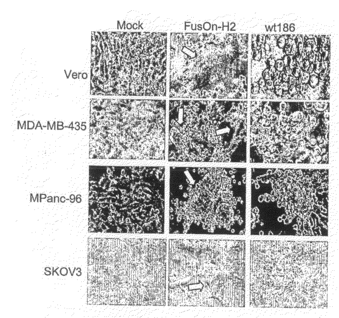
[0173]To determine the phenotype of FusOn-H2, the present inventors infected Vero cells with either wild type HSV-2 or FusOn-H2, or the cells were left uninfected. Twenty-four hours after infection, a clear syncytial formation was visible in the cell monolayer infected with FusOn-H2. No syncytium was seen in either uninfected cells or cells infected with the wild type HSV-2. Similar syncytial formation was also observed in human tumor cells of different tissue origin. In some tumor cells, the infection of wild type HSV-2 also induced some syncytial formation. However, the syncytial formation induced by FusOn-H2 on these cells usually was significantly more profound. So in this case, the FusOn-H2 has an enhanced fusogenic activity when compared with the parental wild type HSV-2. These results indicate that FusOn-H2 is phenotypically different from the parental virus in that its infection induces widespread syncytial formation or enhance...
PUM
| Property | Measurement | Unit |
|---|---|---|
| Weight | aaaaa | aaaaa |
| Weight | aaaaa | aaaaa |
| Weight | aaaaa | aaaaa |
Abstract
Description
Claims
Application Information
 - R&D
- Intellectual Property
- Life Sciences
- Materials
- Tech Scout
- Unparalleled Data Quality
- Higher Quality Content
- 60% Fewer Hallucinations
Browse by: Latest US Patents, China's latest patents, Technical Efficacy Thesaurus, Application Domain, Technology Topic, Popular Technical Reports.
© 2025 PatSnap. All rights reserved.Legal|Privacy policy|Modern Slavery Act Transparency Statement|Sitemap|About US| Contact US: help@patsnap.com


