Method of Treating Dental Periapical Lesions

a technology of periapical lesions and treatment methods, applied in the field of treatment methods of dental periapical lesions, can solve the problems of pulp infection or injury, pain, sensitivity to hot or cold foods, etc., and achieve the effect of promoting healing and slowing down the speed

Inactive Publication Date: 2009-10-15
TOYOTA JIDOSHA KK +1
View PDF29 Cites 26 Cited by
  • Summary
  • Abstract
  • Description
  • Claims
  • Application Information

AI Technical Summary

Benefits of technology

[0016]As will be described more particularly below, since the method of the present invention does not involve cutting through the gums and the jawbone of the patient, it avoids many of the drawbacks of the existing techniques for removing dental periapical lesions, including post operative pain, swelling, possible complications, and risk of infection or injury to nerves, soft tissue, bone and adjacent teeth. In addition, the method of the present invention can be utilized for virtually all dental periapical lesions, even those in teeth which are less accessible using conventional surgical procedures.
[0019]Devices utilizing rotary resecting or ablating filaments are presently preferred. Such devices enable controlled resection or ablation of lesion tissue without damaging surrounding healthy tissues. For example, a high speed rotating biocompatible or biodegradable polymeric filament, composed of, for example, polydioxanone, polylactic acid (PLA) or polyglycolic acid (PGA), can be attached to a dental drill head and inserted via an opening made in the tooth crown into the root canal. When rotated at high speed, the centrifugal force forces the end of the filament to angle outwardly of the filament axis, and to grind away, or ablate, the tissue. An alloy filament made, for example, from Nickel-Titanium (NiTi) can also be used at lower speeds. The alloy can be a pre-shaped shape-memory alloy that has an austenitic final (Af) transition temperature less than body temperature (e.g. 25° C.). Thus, at body temperature, the alloy filament will transition into the austenitic phase and curve outwardly to a predetermined shape such that, when rotated, it will grind and resect or ablate the soft tissue of the lesion, without damaging the surrounding bone tissue.
[0020]Ablation of lesion tissue can be accompanied by resection of the root tip. Such root tip resection is advantageous in that it removes a potential source of infection (e.g. bacteria) present in branches of the main root canal and in the microscopic side canals (dentinal tubules) branching from the main root canal. Resection of the root tip is also advantageous in that it promotes healing.

Problems solved by technology

A deep cavity, a cracked filling, or a cracked tooth can lead to pulp infection or injury.
This in turn can lead to pulp inflammation and infection which may spread to the root canal, often causing sensitivity to hot or cold foods and pain, among other problems.
If not treated at this stage the pulp may then become necrotic and infected.
To prevent that, the host mounts an inflammatory response around the apical foramen which results in local bone destruction.
Periapical lesions may also develop when a previous root canal treatment (as detailed below) was unsuccessful in adequately performing its main task of elimination of bacteria or when prior root canal filling and / or coronal restorations are leaking, thus allowing bacteria to re-contaminate the root canal.
Although widely practiced, apicoectomy is an invasive surgical procedure and as such it is commonly accompanied by postoperative pain, swelling and complications.
In addition, it carries a risk of infection and injury to nerves, soft tissue, bone and adjacent teeth.
Furthermore, some teeth are less accessible or inaccessible surgically (e.g. palatal roots of upper molar), and as such, this procedure cannot be utilized in some periapical lesions.
Finally, this procedure oftentimes results in aesthetic problems such as scarring and recession of gums around restored crown and bridgework.

Method used


Image

  • Method of Treating Dental Periapical Lesions
  • Method of Treating Dental Periapical Lesions
  • Method of Treating Dental Periapical Lesions

Examples


Embodiment Construction

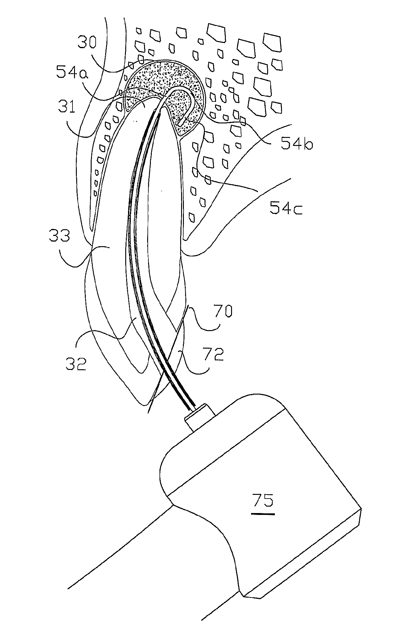
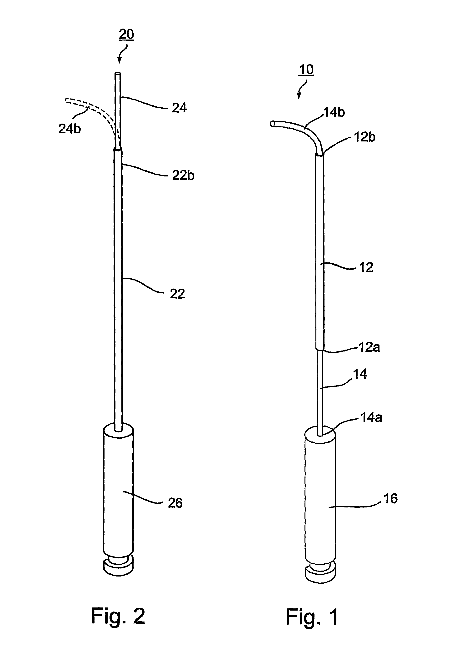
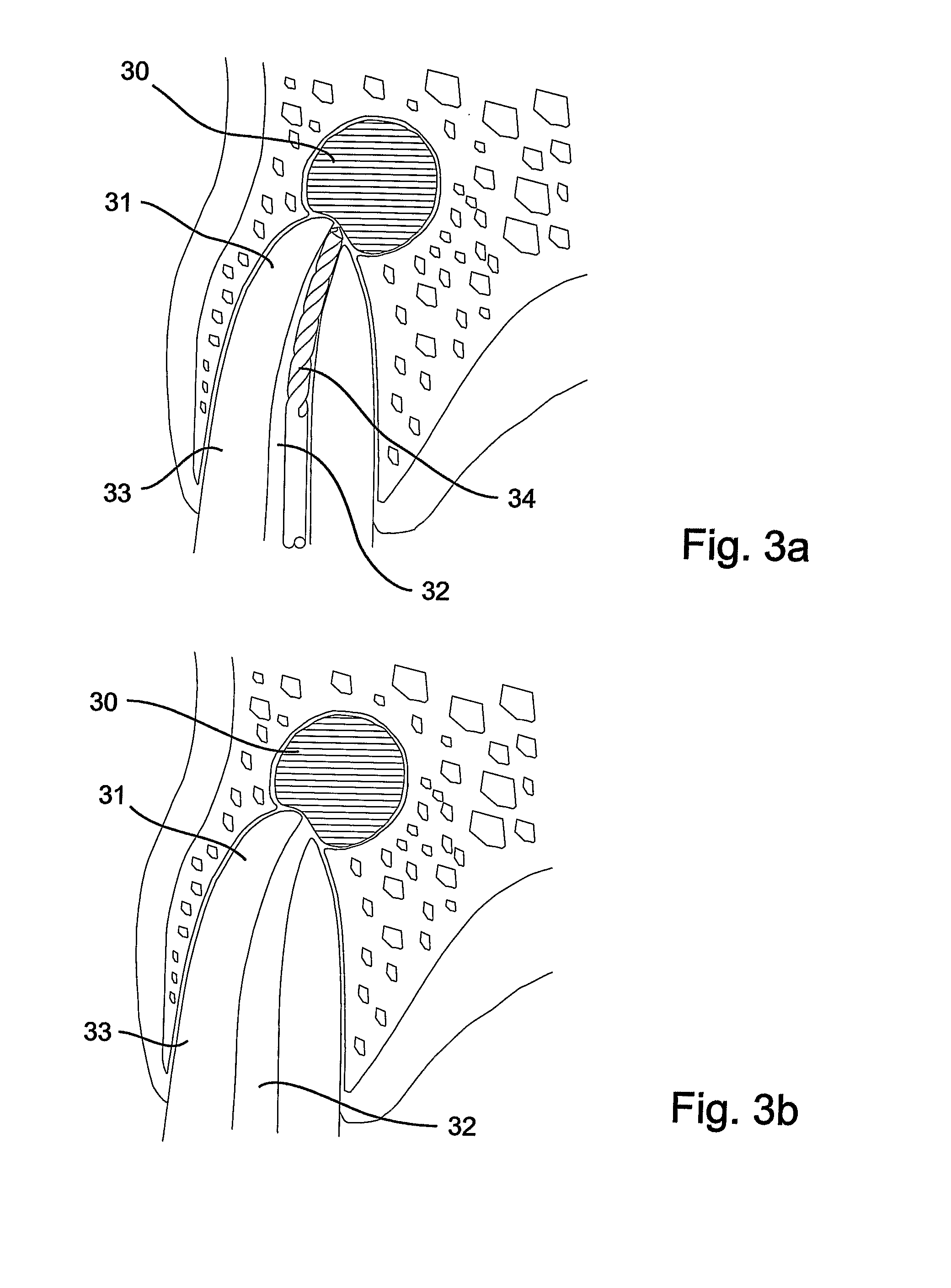
[0039]As indicated earlier, the present invention provides apparatus particularly useful for removing dental periapical lesions at an apex of a root of a tooth. For this purpose, the apparatus provides a rotatable ablating device sized and constructed for (a) introduction through a cavity in the tooth into the root canal; (b) movement therethrough to protrude through the apical foramen into contact with the dental periapical lesion; and (c) rotation while in contact with the dental periapical lesion in order to mince the lesion by ablation so that the particles may be removed via the apical foramen.

[0040]While the invention is particularly useful for removing dental periapical lesions, it can also be used in a wide range of laparoscopic procedures, as well as less invasive subcutaneous and endoscopic procedures. The terms “laparoscopic” and “endoscopic” are interchangeably used herein to refer to surgical procedures performed through small, natural or artificially created openings o...


PUM


Abstract

A method of treating a dental periapical lesion at an apex of a tooth root canal, by accessing the dental periapical lesion via the root canal; and removing the dental periapical lesion via the root canal. In the described preferred embodiments, the dental periapical lesion is accessed via an opening made in the tooth crown leading to the root canal; and the dental periapical lesion is removed by passing a rotary ablating device through the tooth crown, the root canal, and the apex, into engagement with the periapical lesion, rotating the ablating device to ablate the dental periapical lesion, and removing debris produced by the ablation of the periapical lesion via the root canal.

Description

FIELD AND BACKGROUND OF THE INVENTION[0001]The present invention relates to a method of treating dental periapical lesions located at an apex of a root canal of a tooth.[0002]A tooth is composed of a crown and one or more roots which anchor the tooth in a jawbone. The crown, made of enamel and dentin, surrounds a pulp chamber which contains the pulp and extends to the root canal or canals. The root canal opens at the tip of the root (apex) through an opening termed “apical foramen”. A deep cavity, a cracked filling, or a cracked tooth can lead to pulp infection or injury. This in turn can lead to pulp inflammation and infection which may spread to the root canal, often causing sensitivity to hot or cold foods and pain, among other problems. If not treated at this stage the pulp may then become necrotic and infected. Bacteria that exit from the root canal through apical foramen may spread into adjacent or remote tissues. To prevent that, the host mounts an inflammatory response aroun...

Claims


Application Information

Patent Timeline
Patent Type & Authority Applications(United States)
IPC IPC(8): A61C5/02
CPCA61B17/32002A61C5/02A61C3/02A61B17/320725A61C5/40
Inventor HUBER, RONENASCHKENASY, JOELTOBIS, IDAN
Owner TOYOTA JIDOSHA KK
Why Eureka
Try Eureka