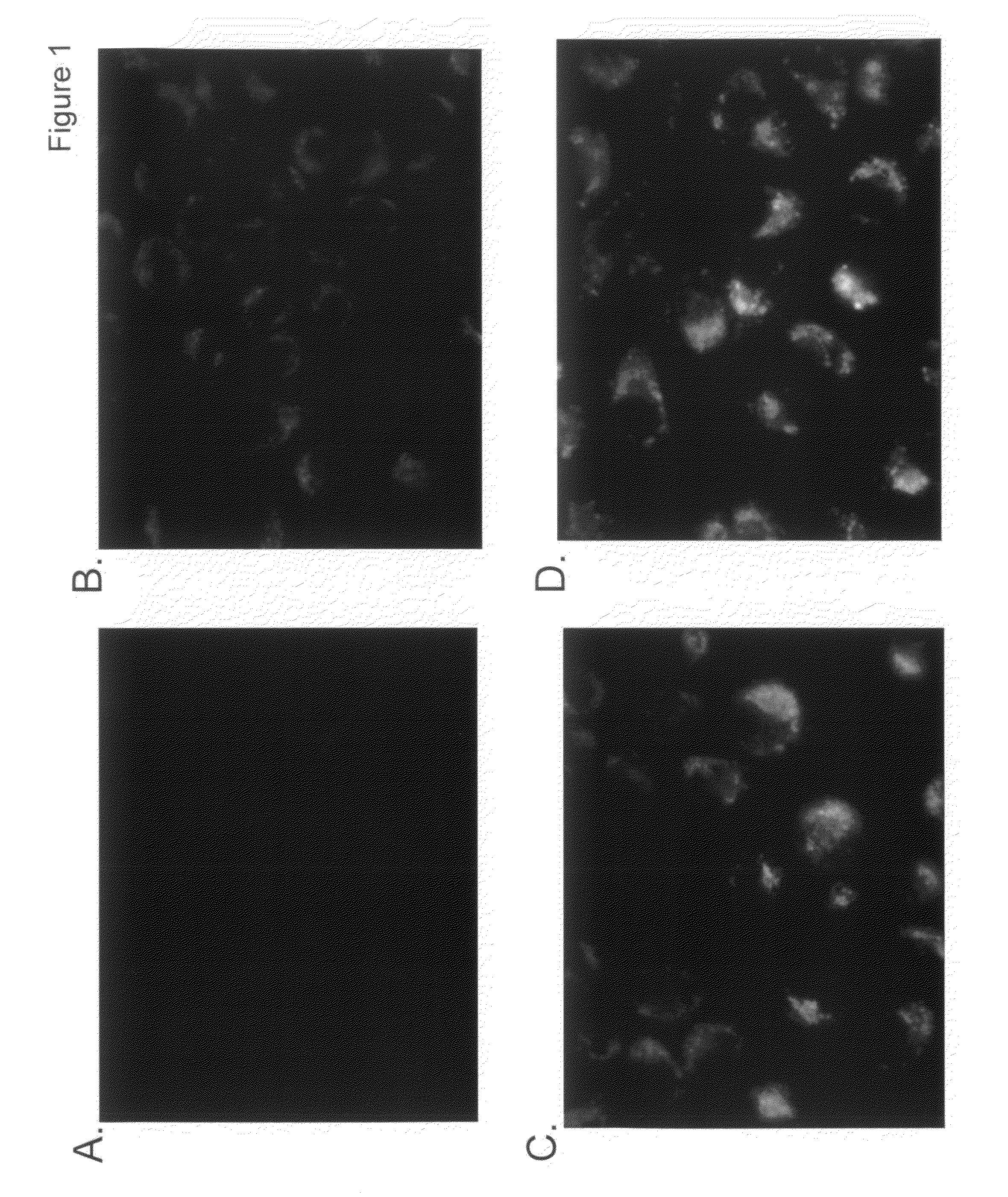
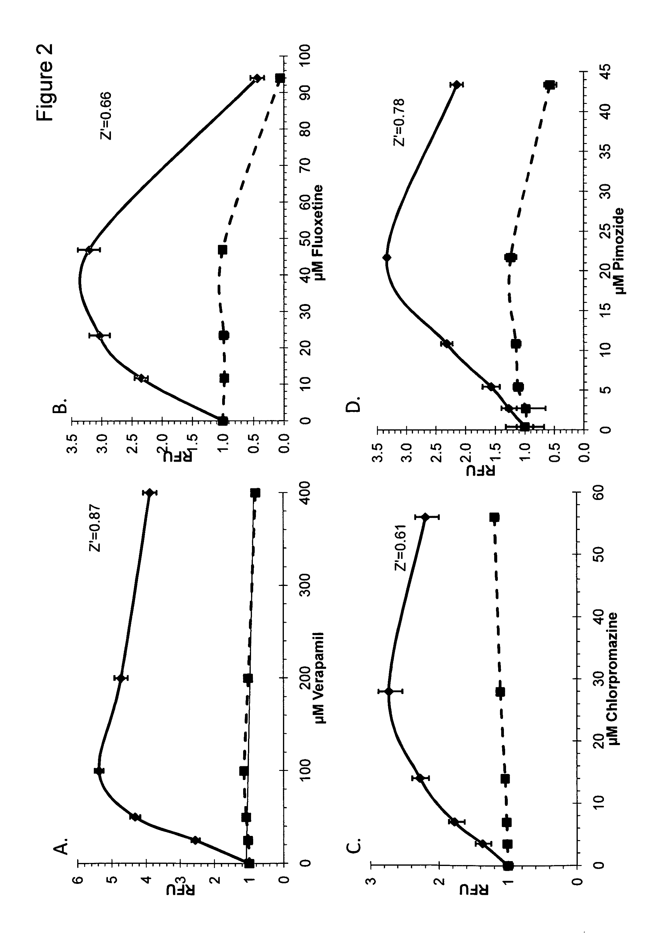
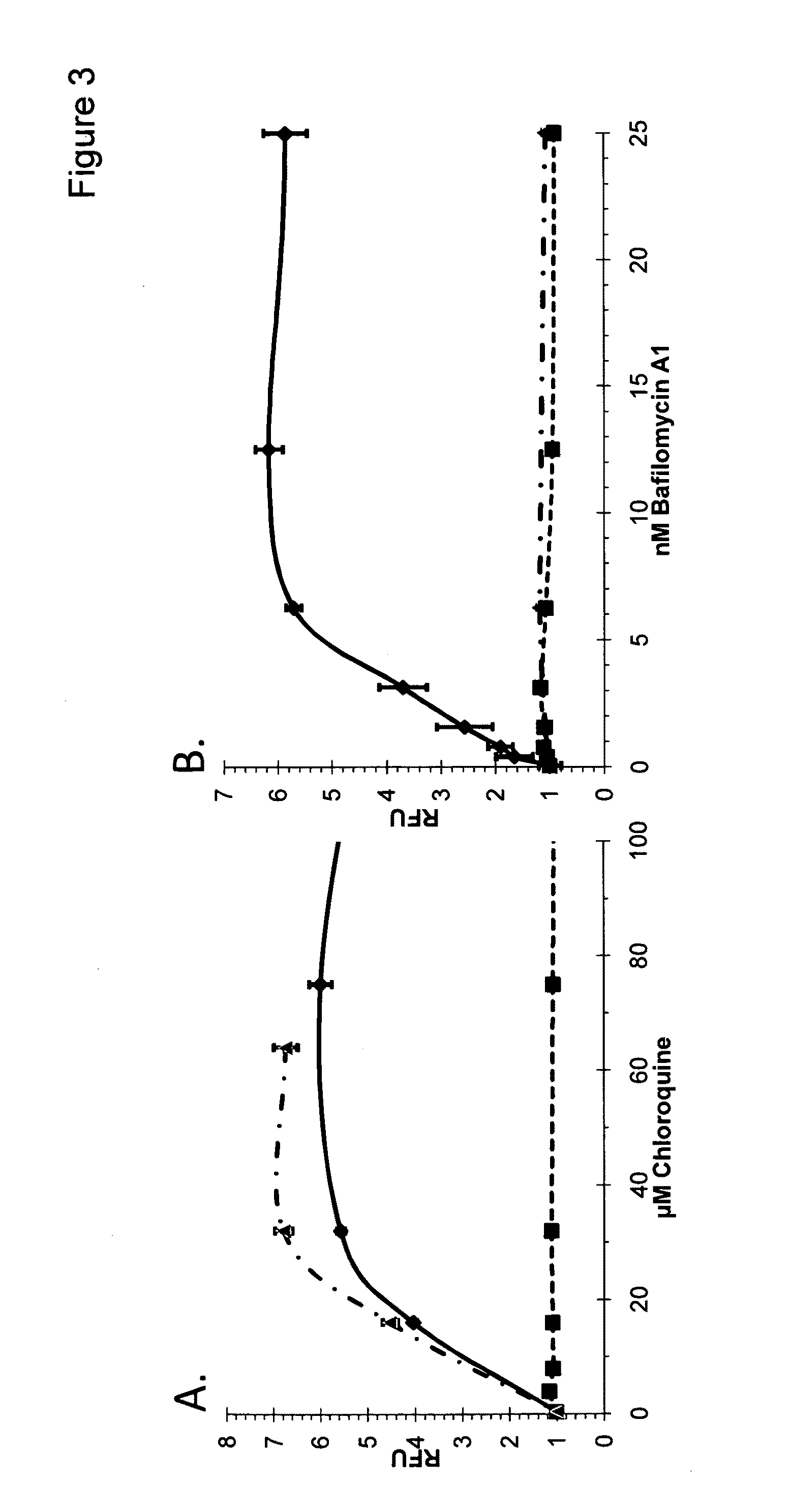
Autophagy and phospholipidosis pathway assays

a phospholipidosis and autophagy technology, applied in the field of lysosomal storage diseases, can solve the problems of shortening the time to market for new drugs, fluorescently-labeled antibodies have few practical applications for intracellular imaging in living cells, and the required additional steps may be time-consuming and expensiv

Active Publication Date: 2010-04-15
ENZO BIOCHEM
View PDF30 Cites 24 Cited by
  • Summary
  • Abstract
  • Description
  • Claims
  • Application Information

AI Technical Summary

Benefits of technology

The technical effect of this patent is an improved methods for testing drugs and other substances for their potential harmfulness to living organisms. This involves creating two samples of cells from different individuals, giving them each a specific test substance, adding a special dye which can be seen inside cells, observing how much of the dye ends up in the cells' empty spaces (vacuoles), and comparing these results between the two samples. If there is no significant difference, it indicates that the substance may have little impact on cells. However, if there is a big change, then we know that the substance could potentially cause damage to cells, leading to possible health risks.

Problems solved by technology

This patent discusses the use of a fluorescent probe called acridine yellow to identify and track changes in membrane structures caused by certain chemicals and diseases like lysosomatosis. However, there are several technical issues related to this method, including interference between different colors, difficulty in obtaining accurate data due to variations in image resolution, and potential danger from high levels of cationic amphiphilic drugs. There is also a lack of effective tools for identifying and measuring phospholipid accumulation during phospholipidosis, making it hard to determine if a specific type of phospholipid is involved without further experimentation. Therefore, the patent seeks to address these challenges and provide a more reliable tool for studying membrane dynamics and disease states.

Method used


Image

  • Autophagy and phospholipidosis pathway assays
  • Autophagy and phospholipidosis pathway assays
  • Autophagy and phospholipidosis pathway assays

Examples


example 1

Synthesis of 1,4-bis(2-(dimethylamino)ethylamino)-2,3-difluoro-5,8-dihydroxyanthracene-9,10-dione (Dye 1)

[0153]A mixture of 1,2,3,4-tetrafluoro-5,8-dihydroxyanthraquinone (1.0 g, 3.2 mmol) and N,N-dimethylethylenediamine (3 mL) in CH2Cl2 (30 mL) was stirred at room temperature for 12 hours. After evaporation of the solvents, the residue was purified by silica gel chromatography using isocratic solvent system of EtOAc / MeOH / Et3N (10:10:1) yielding 830 mgs of Dye 1 as dark blue product. Abs (max, PBS pH 7.4)=568 nm; Em=675 nm. The structure of Dye 1 is given below:

example 2

Synthesis of 1,4-bis(5-(diethylamino)pentan-2-ylamino)-2,3-difluoro-5,8-dihydroxyanthracene-9,10-dione (Dye 2)

[0154]A mixture of 1,2,3,4-tetrafluoro-5,8-dihydroxyanthraquinone (0.47 g, 1.5 mmol) and 2-Amino-5-diethylaminopentane (2.3 mL) in CH2Cl2 (20 mL) was stirred at room temperature for 12 hours. After evaporation of the solvents, the residue was purified by silica gel chromatography using isocratic solvent system of CH2Cl2 / MeOH / Et3N (100:10:1) yielding 364 mgs of Dye 2 as dark blue product. Abs (max, PBS pH 7.4)=667 nm; Em=700 nm. The structure of Dye 2 is given below:

example 3

Synthesis of Dye 3

(a) Preparation of 2-((naphthalene-1-yloxy)methyl)oxirane (Compound 1)

[0155]To a solution of 1-naphthol (3.0 g, 0.02 mol) in DMSO (10 mL), potassium hydroxy (flakes, 2.0 g, 0.06 mol) was added. The combined mixture was stirred at room temperature for 30 min and then epichlorohydrin (5.6 g, 4.7 mL, 0.06 mol) was added slowly over a period of 45 min and stirring was continued for 16 hours. The reaction was quenched with water (30 mL) and extracted with chloroform (2×50 mL). The combined organic layers were washed with 1N aqueous NaOH (2×50 mL), water (2×100 mL) and brine (2×100 mL) and dried over MgSO4. The solvent was removed under reduced pressure to provide Compound 1 as a yellow liquid (3.26 g, 78%). This product was used without further purification. The structure of Compound 1 is given below:

(b) Preparation of 1-(3-ammoniopropyl)-4-methylquinolinium bromide (Compound 2)

[0156]A mixture of lepidine (3.0 g, 21 mmol) and 3-bromopropylamine hydrobromide (4.6 g, 21 m...


PUM


Abstract

Provided are assays useful for detecting and monitoring autophagy and phospholipidosis, including the progression of lysosomal storage diseases. Drugs and treatments for lysosomal storage diseases can be monitored for effectiveness in lysosomal storage disease conditions. Drug candidates and suspected toxic agents can also be screened for toxicity to cells, tissues and organs. Also provided are methods for distinguishing between phospholipidosis activators and autophagy pathway perturbation agents.

Description


Claims


Application Information

Patent Timeline
Owner ENZO BIOCHEM
Why Eureka
Try Eureka