Endoscope system

a technology of endoscope and endoscope, which is applied in the direction of optical radiation measurement, instruments, catheters, etc., can solve the problems of difficult to obtain the distribution image of each fluorescent agent, low diagnostic accuracy, etc., and achieve the effect of improving diagnostic accuracy of cancer cells

Active Publication Date: 2010-04-15
OLYMPUS CORP
View PDF12 Cites 40 Cited by
  • Summary
  • Abstract
  • Description
  • Claims
  • Application Information

AI Technical Summary

Benefits of technology

[0016]According to the present invention, a fluorescence distribution image of each fluorescent agent can be acquired from a fluorescence image that has been captured in a mixed-color state, without using a special apparatus such as a variable spectral element, and the diagnosability of cancer cells can be improved.

Problems solved by technology

Therefore, the diagnosability for specifying cancer cells tends to be low only with a single type of fluorescent probe.
Therefore, it is difficult to obtain the distribution image of each fluorescent agent.

Method used


Image

  • Endoscope system
  • Endoscope system
  • Endoscope system

Examples


Embodiment Construction

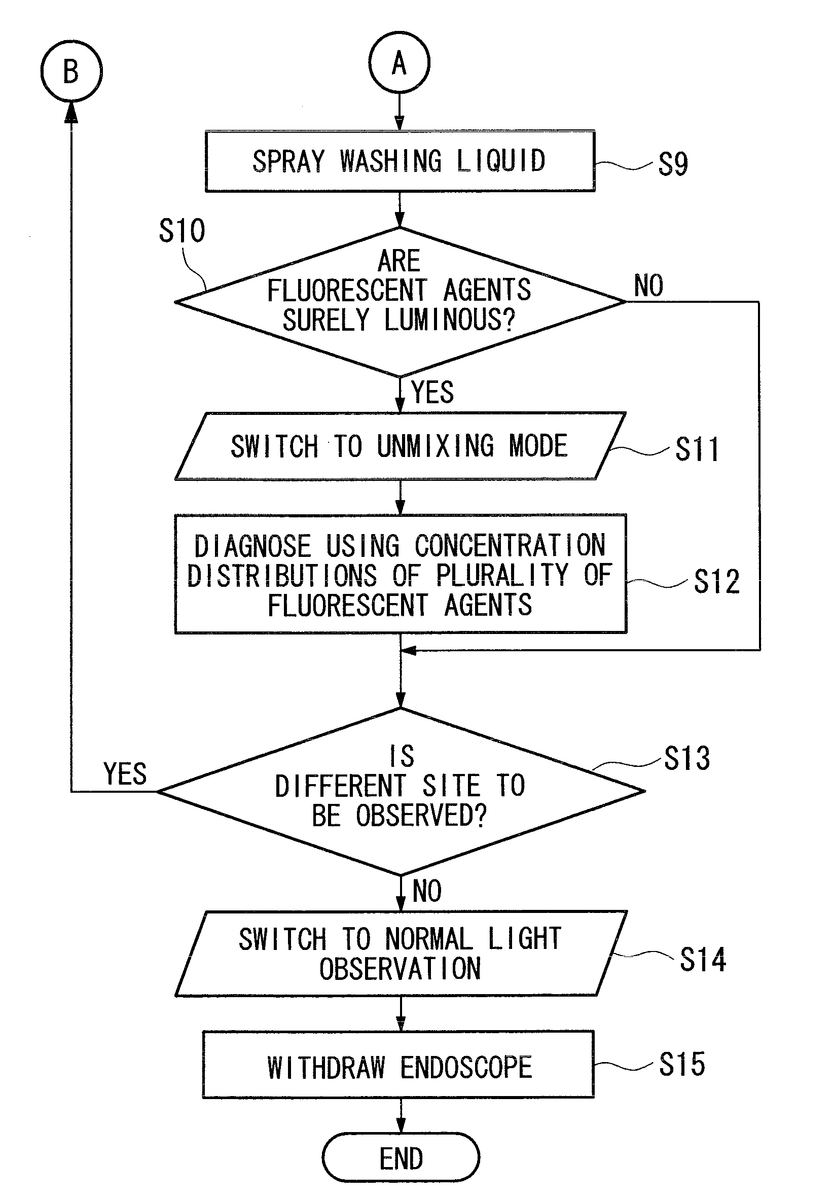
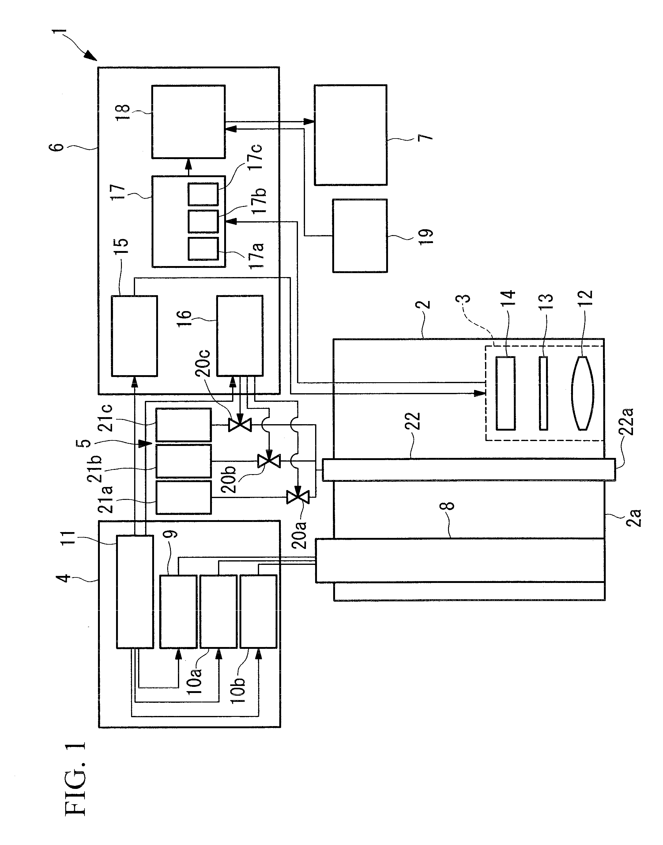
[0029]Hereunder is a description of an endoscope system 1 according to a first embodiment of the present invention, with reference to FIG. 1 to FIG. 5.

[0030]As shown in FIG. 1, the endoscope system 1 according to this embodiment comprises an insertion section 2 to be inserted into a body cavity of an organism, an imaging unit (imaging section) 3 disposed in the insertion section 2, a light source unit (light source section) 4 for emitting excitation light and illumination light for normal light observation, a liquid delivery unit 5 for supplying a liquid to be discharged from the distal end 2a of the insertion section 2, a control unit 6 for controlling the imaging unit 3, the light source unit 4, and the liquid delivery unit 5, and a display unit (display section) 7 for displaying the image captured by the imaging unit 3.

[0031]The insertion section 2 has an extremely narrow outer dimension to be insertable into a body cavity of an organism, and comprises therein a light guide 8 for...


PUM


Abstract

A fluorescence distribution image of each fluorescent agent can be acquired from a fluorescence image that has been captured in a mixed state so as to improve the diagnosability of cancer cells. An endoscope system comprises: a light source section for selectively irradiating two or more types of excitation lights having different spectral characteristics so as to excite two or more types of fluorescent agents having different optical characteristics; an imaging section provided on a part to be inserted into a body cavity, and having a light-receiving sensitivity in the wavelength bands of the two or more types of fluorescence radiated from the observation target by respective types of excitation lights; a storage section for storing relation information between the fluorescence intensity and the concentration of each of the fluorescent agents; a concentration information computing section for computing concentration information of each of the fluorescent agents on the basis of the fluorescence intensities of two or more images captured by the imaging section and the relation information stored in the storage section, and outputting the concentration information; and a mode switching section capable of switching between a first fluorescence observation mode in which at least one of the fluorescence intensity images acquired by the imaging section is presented, and a second fluorescence observation mode in which the concentration information of each of the fluorescent agents computed by the concentration information computing section is presented.

Description

TECHNICAL FIELD [0001]The present invention relates to an endoscope system.BACKGROUND ART [0002]Conventionally, it is known that some types of proteins etc. are overexpressed in cancer cells, as compared to in normal sites. A method has been proposed in which a fluorescent probe is used to make thus expressed proteins or other molecules luminous to thereby diagnose cancer cells, as well as observing this fluorescence in an endoscopic manner to thereby identify these cancer cells (for example, refer to Patent Document 1).[0003]In Patent Document 1, an endoscope apparatus in which a single type of fluorescent probe is used for diagnosing cancer cells, has been disclosed.[0004]Patent Document 1:[0005]Japanese Unexamined Patent Application, Publication No. Hei 10-201707DISCLOSURE OF INVENTION [0006]However, even in non-cancerous sites such as a site of inflammation occurrence do exist molecules expressed by cancer cells, which may emit similar fluorescence. Therefore, the diagnosability...

Claims


Application Information

Patent Timeline
Patent Type & Authority Applications(United States)
IPC IPC(8): A61B1/06A61B6/00
CPCA61B1/043A61B1/05A61B1/0638A61B5/0071G01N2021/6441G01J3/2823G01J3/4406G01N21/6456A61B5/0084A61B1/0655
Inventor NAKAOKA, MASAYAMORISHITA, KOKI
Owner OLYMPUS CORP
Why Eureka
Try Eureka