Method for reconstructing images of an imaged subject from a parallel MRI acquisition

Active Publication Date: 2010-12-09
SIEMENS HEALTHCARE GMBH +1
View PDF6 Cites 34 Cited by
  • Summary
  • Abstract
  • Description
  • Claims
  • Application Information

AI Technical Summary

Benefits of technology

[0008]The above problems are obviated by the present invention which provides a method for reconstructing images of an imaged subject from a parallel MRI acquisition, comprising generating, for a response signal received by a respective RF reception coil, a complete dataset in k-space from a reduced k-space dataset of the response signal and associated weighting factors for the reduced k-space dataset; spatially transforming the complete k-space dataset generated to obtain an estimated reconstructed image for a respective RF reception coil; maximizing the sparsity of the estimated reconstructed images; repeating the generating and the spatially transforming steps using revised associated weighting factors obtained from each respective k-space dataset that results from the maximizing step; and combining the estimated reconstructed images for the respective RF reception coils to obtain a reconstructed image of the imaged subject. The generating step and the spatially transforming step may comprise using a generalized autocalibrating partially parallel acquisitions technique. In such case, or separately, the maximizing step may comprise using a compressed sensing technique. Also separately, the generating step may comprise deriving the associated weighting factors from reference calibration data for a respective reduced k-space dataset. Alternatively, the generating step may comprise deriving the associated weighting factors from reference calibration data that is obtained from a respective reduced k-space dataset. The generating step may comprise deriving the associated weighting factors from reference calibration data that is obtained from a respective reduced k-space dataset and the repeating step may comprise deriving the revised associated weighting factors from reference calibration data that is obtained from a respective complete k-space dataset that results from the maximizing step. Further, the maximizing step may comprise optimizing a sparsifying transform of an estimated reconstructed image over the set of values of the unacquired k-space data missing in the respective reduced k-space dataset. In such case, the generating step may comprise deriving the associated weighting factors from reference calibration data that is obtained from a respective reduced k-space dataset and the repeating step may comprise deriving the revised associated weighting factors from reference calibration data that is obtained from a respective complete k-space dataset that results from the maximizing step. Also, the maximizing step may comprise using a compressed sensing technique and may further comprise optimizing a sparsifying transform of an estimated reconstructed image over the set of values of the unacquired k-space data missing in the respective reduced k-space dataset. Also, the combining step may comprise combining the estimated reconstructed images using a sum of squares reconstruction to obtain a reconstructed image of the imaged subject.

Problems solved by technology

However, there is a fundamental trade-off in MRI between acquisition time and image quality of the reconstructed images.
This technique results in a significant decrease in the acquisition time, allowing for shorter examinations or for higher temporal sampling rates in dynamic imaging.

Method used


Image

  • Method for reconstructing images of an imaged subject from a parallel MRI acquisition
  • Method for reconstructing images of an imaged subject from a parallel MRI acquisition
  • Method for reconstructing images of an imaged subject from a parallel MRI acquisition

Examples


Embodiment Construction

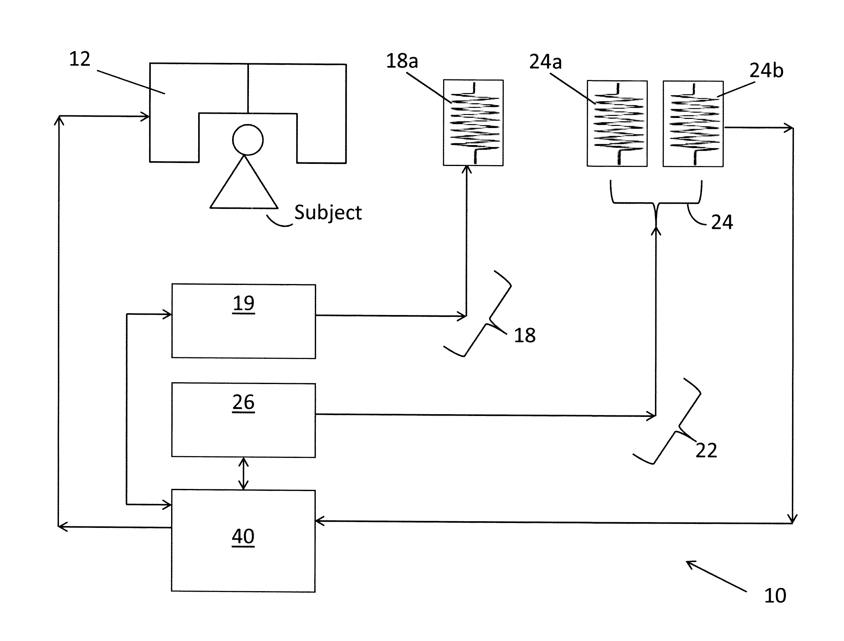
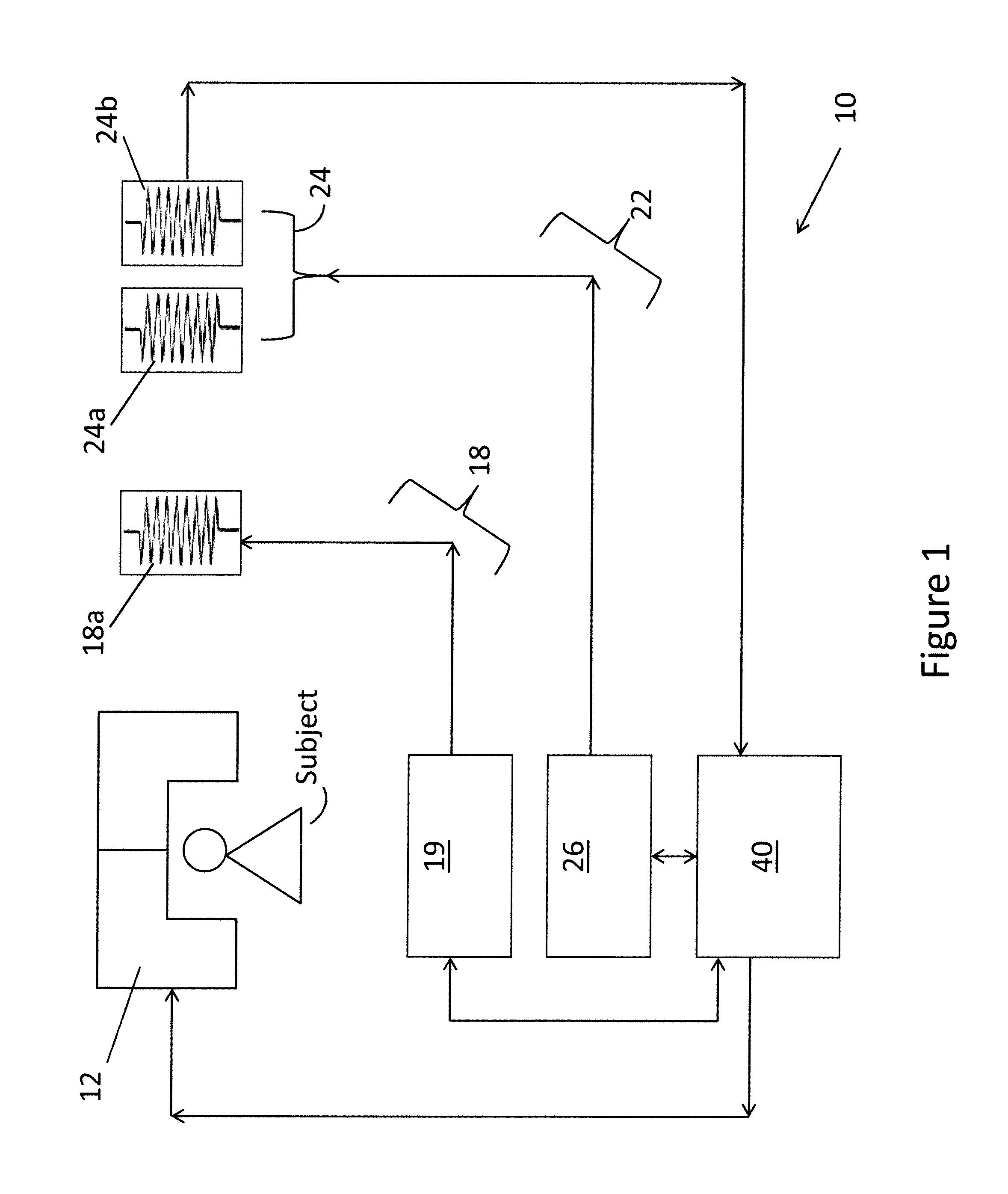
[0016]FIG. 1 is a block diagram of a conventional MRI scanner 10 (simplified) that performs parallel MR image reconstruction in accordance with the present invention. A main magnet 12 generates a strong static magnetic field in an imaging region where the subject (i.e., patient or the specific body part of a patient to be examined) is introduced. The magnet 12 is used to polarize the patient's body part, i.e., certain atoms in the patient's body part that were previously randomly-ordered become aligned along the magnetic field. A gradient coil system 18, having a gradient coil subsystem 18a and a gradient coil control unit 19, generates a time-varying linear magnetic field gradient in respective spatial directions, x, y and z, and spatially encodes the positions of the polarized or excited atoms. An RF system 22, having an RF coil subsystem 24 and a pulse generation unit 26, transmits a series of RF pulses to the patient's body part to excite the “ordered” atoms of the patient's bod...


PUM


Abstract

A parallel MR imaging method that uses a reconstruction algorithm that combines the GRAPPA image reconstruction method and the compressed sensing (CS) image reconstruction method in an iterative approach (200) or joint energy optimization approach (300).

Description

CROSS-REFERENCE TO RELATED APPLICATIONS[0001]This application claims the benefit of Provisional U.S. Patent Application Ser. No. 61 / 181,312 entitled, “System For Reconstructing Images From A Parallel MRI Acquisition Via Combined GRAPPA And Compressed Sensing”, filed in the name of Leo Grady and Johnathan R. Polimeni on May 27, 2009, the disclosure of which is also hereby incorporated herein by reference.FIELD OF INVENTION[0002]The present invention relates to magnetic resonance imaging (MRI). More particularly, the present invention relates to parallel MRI image reconstruction.BACKGROUND OF THE INVENTION[0003]Magnetic resonance imaging (MRI) is a non-invasive diagnostic imaging procedure that uses nuclear magnetization and radio waves to produce internal images of a patient. The operation of an MRI scanner is well known but, briefly, an MRI scanner contains magnetic coils that create a strong static magnetic field in which the patient is positioned. Certain atoms in a patient's body...

Claims


Application Information

Patent Timeline
IPC IPC(8): G01R33/48
CPCG01R33/5611
Inventor GRADY, LEOPOLIMENI, JONATHAN R.
Owner SIEMENS HEALTHCARE GMBH
Why Eureka
Try Eureka