System for Accelerated MR Image Reconstruction

a mri image and image reconstruction technology, applied in the field of parallel mri image reconstruction, can solve the problems of spir-it being more computationally intensive, affecting the accuracy of mri reconstruction, and reducing the acquisition time, so as to maximize the sparsity of estimated reconstructed images

Active Publication Date: 2012-04-05
SIEMENS HEALTHCARE GMBH +1
View PDF4 Cites 42 Cited by
  • Summary
  • Abstract
  • Description
  • Claims
  • Application Information

AI Technical Summary

Benefits of technology

[0010]The above problems are obviated by the present invention which provides a method for reconstructing images of an imaged subject from a parallel MRI acquisition, comprising generating, for a response signal received by a respective RF reception coil, a complete dataset in k-space from a reduced k-space dataset of the response signal and associated weighting factors for the reduced k-space dataset; spatially transforming the complete k-space dataset generated to obtain an estimated reconstructed image for a respective RF reception coil; maximizing the sparsity of the estimated reconstructed images; repeating the generating and the spatially transforming steps using revised associated weighting factors obtained from each respective k-space dataset that results from the maximizing step; and combining the estimated reconstructed images for the respective RF reception coils to obtain a reconstructed image of the imaged subject. The generating step and the spatially transforming step may comprise using a generalized autocalibrating partially parallel acquisitions technique. In such case, or separately, the maximizing step may comprise using a compressed sensing technique. Also separately, the generating step may comprise deriving the associated weighting factors from reference calibration data for a respective reduced k-space dataset. Alternatively, the generating step may comprise deriving the associated weighting factors from reference calibration data that is obtained from a respective reduced k-space dataset. The generating step may comprise deriving the associated weighting factors from reference calibration data that is obtained from a respective reduced k-space dataset and the repeating step may comprise deriving the revised associated weighting factors from reference calibration data that is obtained from a respective complete k-space dataset that results from the maximizing step. Further, the maximizing step may comprise optimizing a sparsifying transform of an estimated reconstructed image over the set of values of the unacquired k-space data missing in the respective reduced k-space dataset. In such case, the generating step may comprise deriving the associated weighting factors from reference calibration data that is obtained from a respective reduced k-space dataset and the repeating step may comprise deriving the revised associated weighting factors from reference calibration data that is obtained from a respective complete k-space dataset that results from the maximizing step. Also, the maximizing step may comprise using a compressed sensing technique and may further comprise optimizing a sparsifying transform of an estimated reconstructed image over the set of values of the unacquired k-space data missing in the respective reduced k-space dataset. Also, the combining step may comprise combining the estimated reconstructed images using a sum of squares reconstruction to obtain a reconstructed image of the imaged subject.
[0011]The present invention may also provide a method for obtaining an image of a subject or sample by a parallel MR imaging device, comprising a) generating image reconstruction data from a generalized autocalibrating partially parallel acquisitions reconstruction of weighted under-sampled k-space data acquired by each RF reception coil of the device; b) calculating new weightings for respective under-sampled k-space data from k-space data obtained from a compressed sensing reconstruction of the image reconstruction data previously generated; and c) reconstructing the image according to the image reconstruction data generated from repeating the generating and calculating steps until convergence is achieved. In such case, the generating step may comprise deriving the weightings from calibration data that is obtained from respective under-sampled k-space data. Also, the compressed sensing reconstruction may obtain k-space data by maximizing the sparsity of a reconstructed image from the image reconstruction data generated. Alternatively, the compressed sensing reconstruction may obtain k-space data by optimizing a sparsifying transform of a reconstructed image over the set of values of the unacquired k-space data missing in the respective under-sampled k-space data. Also, the reconstructing step may comprise reconstructing using a weighted combination of the individual image reconstructions from each RF reception coil of the device.
[0014]An MR imaging system uses the multiple RF coils for acquiring corresponding multiple image data sets of the slice or volume. An image data processor comprises at least one processing device conditioned for generating a composite MR image data set representing a single image in a single non-iterative operation by performing a weighted combination of luminance representative data of individual corresponding pixels of the multiple image data sets in providing an individual pixel luminance value of the composite MR image data set. The image data processor reduces noise in the composite MR image data set by generating a reduced set of significant components in a predetermined transform domain representation of data representing the composite image to provide a de-noised composite MR image data set. An image generator comprises at least one processing device conditioned for generating a composite MR image using the de-noised composite MR image data set.

Problems solved by technology

However, there is a fundamental trade-off in MRI between acquisition time and image quality of the reconstructed images.
This technique results in a significant decrease in the acquisition time, allowing for shorter examinations or for higher temporal sampling rates in dynamic imaging.
The known systems reconstruct diagnostically useful images from undersampled data, however, at higher acceleration factors, GRAPPA especially tends to fail due to noise enhancement and unresolved aliasing.
Also, SPIR-iT is more computationally intensive than GRAPPA for uniform undersampling.
However, the combination of these two frameworks remains not fully explored.
CS-GRAPPA, CS-SENSE, and L1 SPIR-iT combine parallel imaging methods (GRAPPA, SENSE, and SPIR-iT) with compressed sensing, however these combinations have substantial limitations.
However, L1 SPIR-iT is computationally complex because the SPIR-IT kernel, unlike the GRAPPA kernel it replaces, needs to be applied repeatedly throughout the process, updating the k-space data for consistency instead of filling in the missing k-space data directly.

Method used


Image

  • System for Accelerated MR Image Reconstruction
  • System for Accelerated MR Image Reconstruction
  • System for Accelerated MR Image Reconstruction

Examples


Embodiment Construction

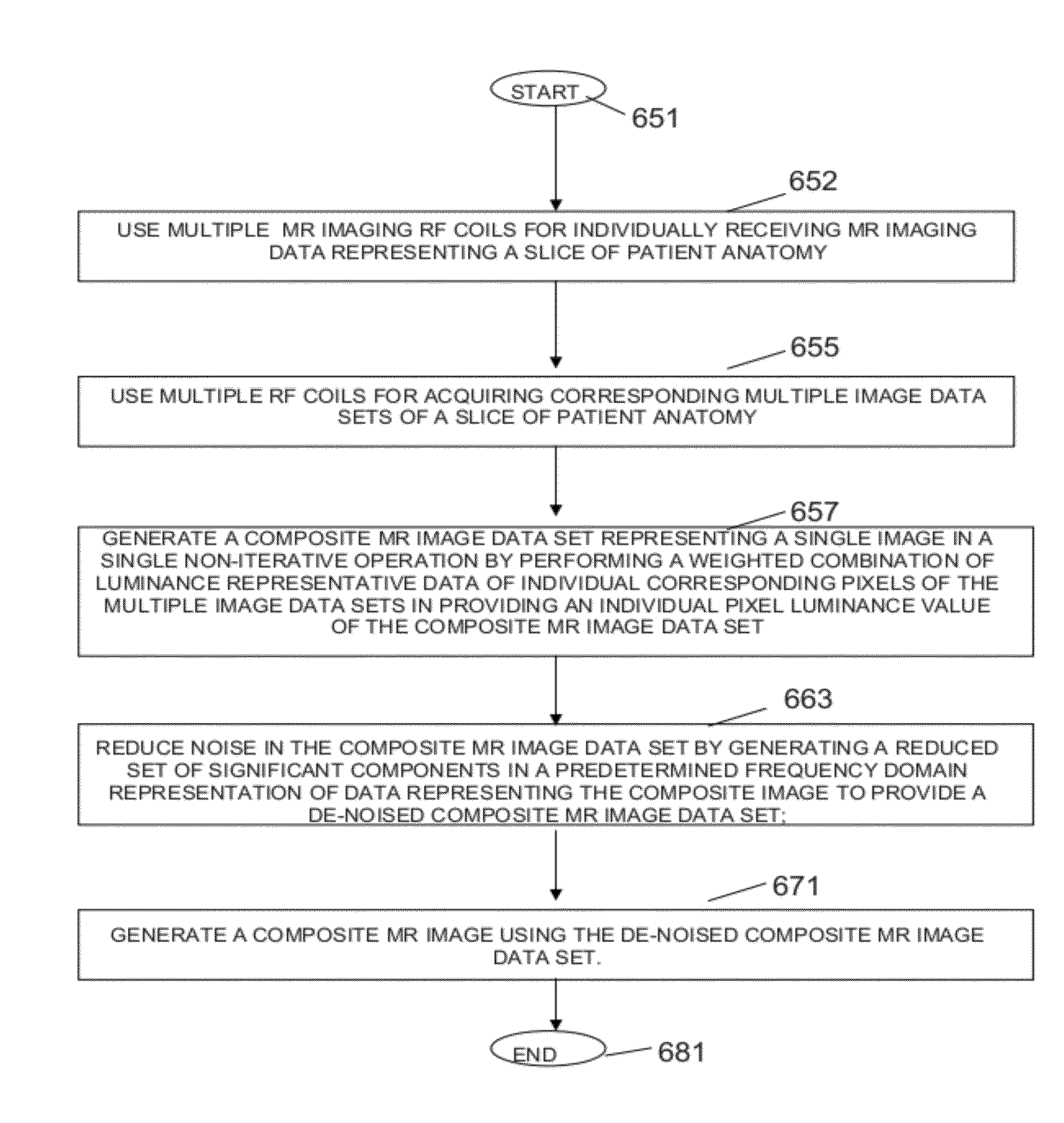
[0029]A system accelerates magnetic resonance (MR) imaging beyond what is achievable using parallel imaging with GRAPPA (Generalized Autocalibrating Partially Parallel Acquisition) or compressed sensing (CS) alone using Sparse Reconstruction of Images employing a Nullspace method and GRAPPA (SpRING). The system adaptively selects an appropriate sparsifying transform and penalty function, and evaluates SpRING performance. The system optimizes trade-off between fidelity to a GRAPPA function and sparsity for different levels of acceleration.

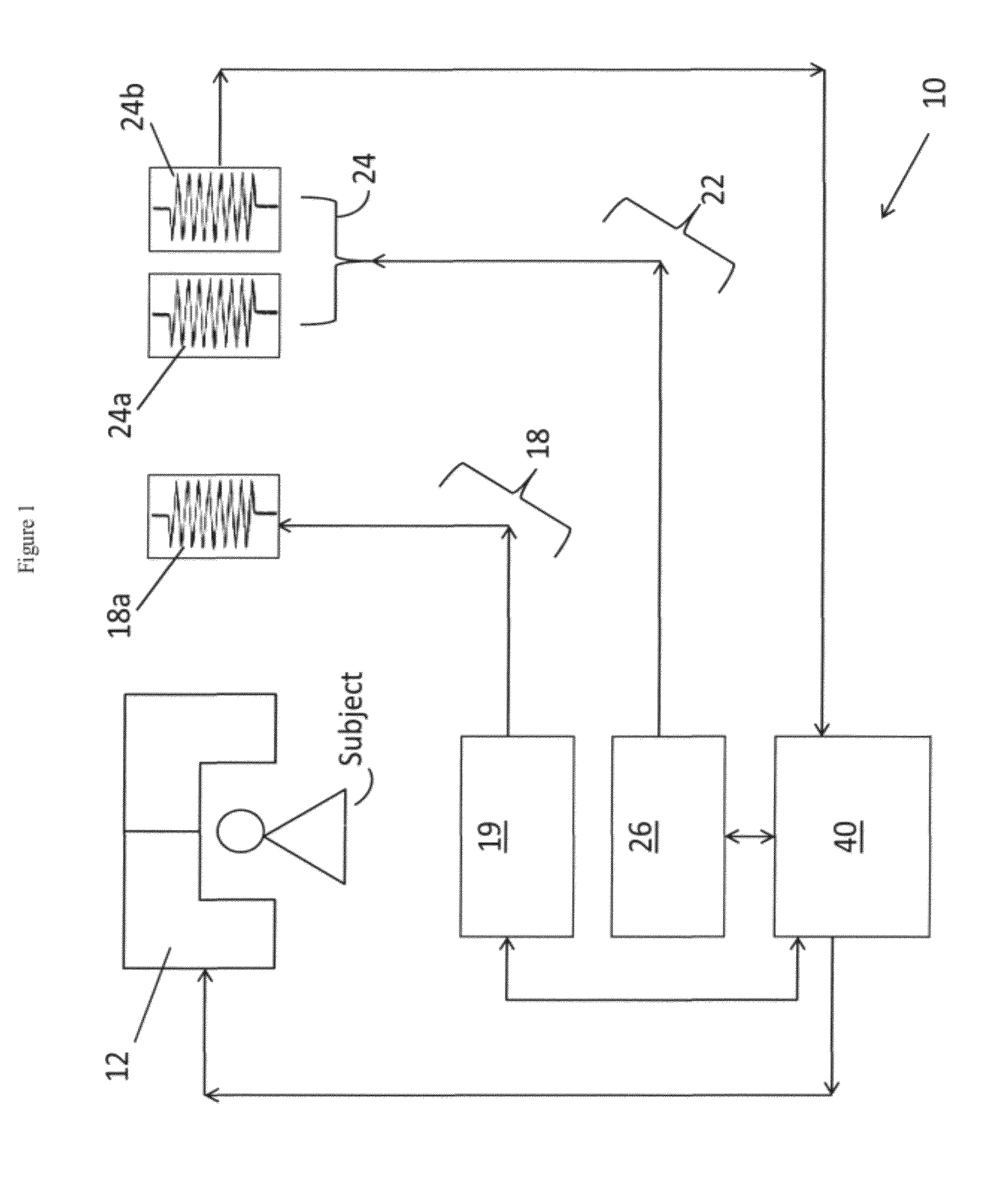
[0030]FIG. 1 is a block diagram of a conventional MRI scanner 10 (simplified) that performs parallel MR image reconstruction in accordance with the present invention. A main magnet 12 generates a strong static magnetic field in an imaging region where the subject (i.e., patient or the specific body part of a patient to be examined) is introduced. The magnet 12 is used to polarize the patient's body part, i.e., certain atoms in the patient's body par...


PUM


Abstract

An MR imaging system uses the multiple RF coils for acquiring corresponding multiple image data sets of the slice. An image data processor comprises at least one processing device conditioned for, generating a composite MR image data set representing a single image in a single non-iterative operation by performing a weighted combination of luminance representative data of individual corresponding pixels of the multiple image data sets in providing an individual pixel luminance value of the composite MR image data set. The image data processor reduces noise in the composite MR image data set by generating a reduced set of significant components in a predetermined transform domain representation of data representing the composite image to provide a de-noised composite MR image data set. An image generator comprises at least one processing device conditioned for, generating a composite MR image using the de-noised composite MR image data set.

Description

[0001]This application claims priority of U.S. Patent Application 61 / 451,162 filed Mar. 10, 2011 by L. Grady et al. and is a continuation-in-part application of U.S. patent application Ser. No. 12 / 788,439 filed 27 May 2010 by L. Grady et al. claiming priority of Provisional U.S. Patent Application Ser. No. 61 / 181,312 filed 27 May 2009 by L. Grady et al.FIELD OF INVENTION[0002]The present invention relates to magnetic resonance imaging (MRI). More particularly, the present invention relates to parallel MRI image reconstruction.BACKGROUND OF THE INVENTION[0003]Magnetic resonance imaging (MRI) is a non-invasive diagnostic imaging procedure that uses nuclear magnetization and radio waves to produce internal images of a patient. The operation of an MRI scanner is well known but, briefly, an MRI scanner contains magnetic coils that create a strong static magnetic field in which the patient is positioned. Certain atoms in a patient's body that were previously randomly-ordered become aligne...

Claims


Application Information

Patent Timeline
Patent Type & Authority Applications(United States)
IPC IPC(8): G01R33/56G01R33/34
CPCG01R33/5611
Inventor WELLER, DANIELGOYAL, VIVEK K.POLIMENI, JONATHAN RIZZOGRADY, LEO
Owner SIEMENS HEALTHCARE GMBH
Why Eureka
Try Eureka