Context driven image mining to generate image-based biomarkers

Active Publication Date: 2011-05-26
DEFINIENS AG
View PDF59 Cites 42 Cited by
  • Summary
  • Abstract
  • Description
  • Claims
  • Application Information

AI Technical Summary

Problems solved by technology

One of the more difficult tasks of detecting patterns in medical images involves detecting cancerous mass lesions and micro-calcifications in X-ray images of breasts (also called mammograms).
Detecting cancerous regions in breasts is made more difficult, however, by the similar appearance of pixels associated with benign and malignant lesions and micro-calcifications.
As the probability threshold o

Method used


Image

  • Context driven image mining to generate image-based biomarkers
  • Context driven image mining to generate image-based biomarkers
  • Context driven image mining to generate image-based biomarkers

Examples


Embodiment Construction

[0060]Reference will now be made in detail to some embodiments of the invention, examples of which are illustrated in the accompanying drawings.

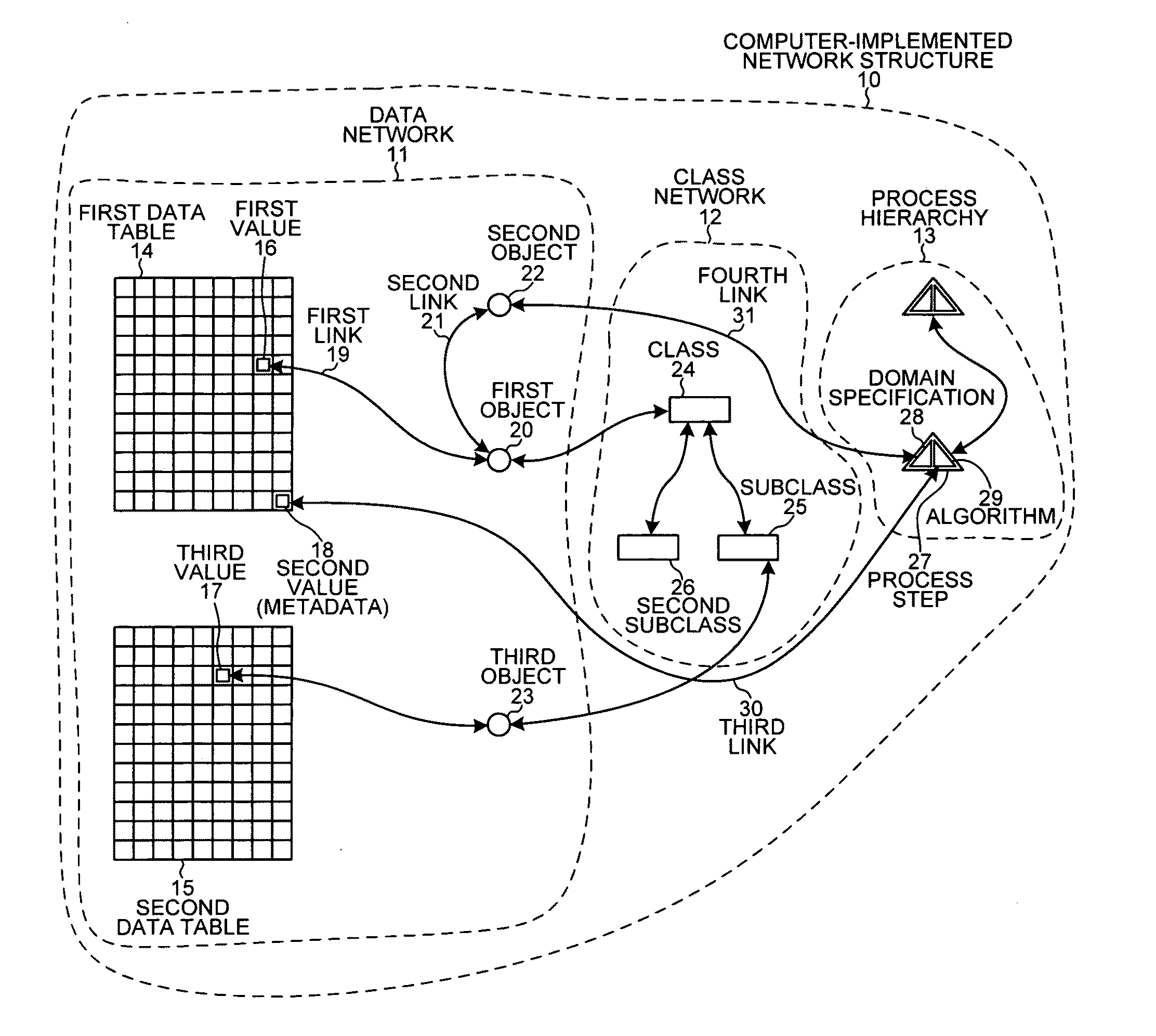
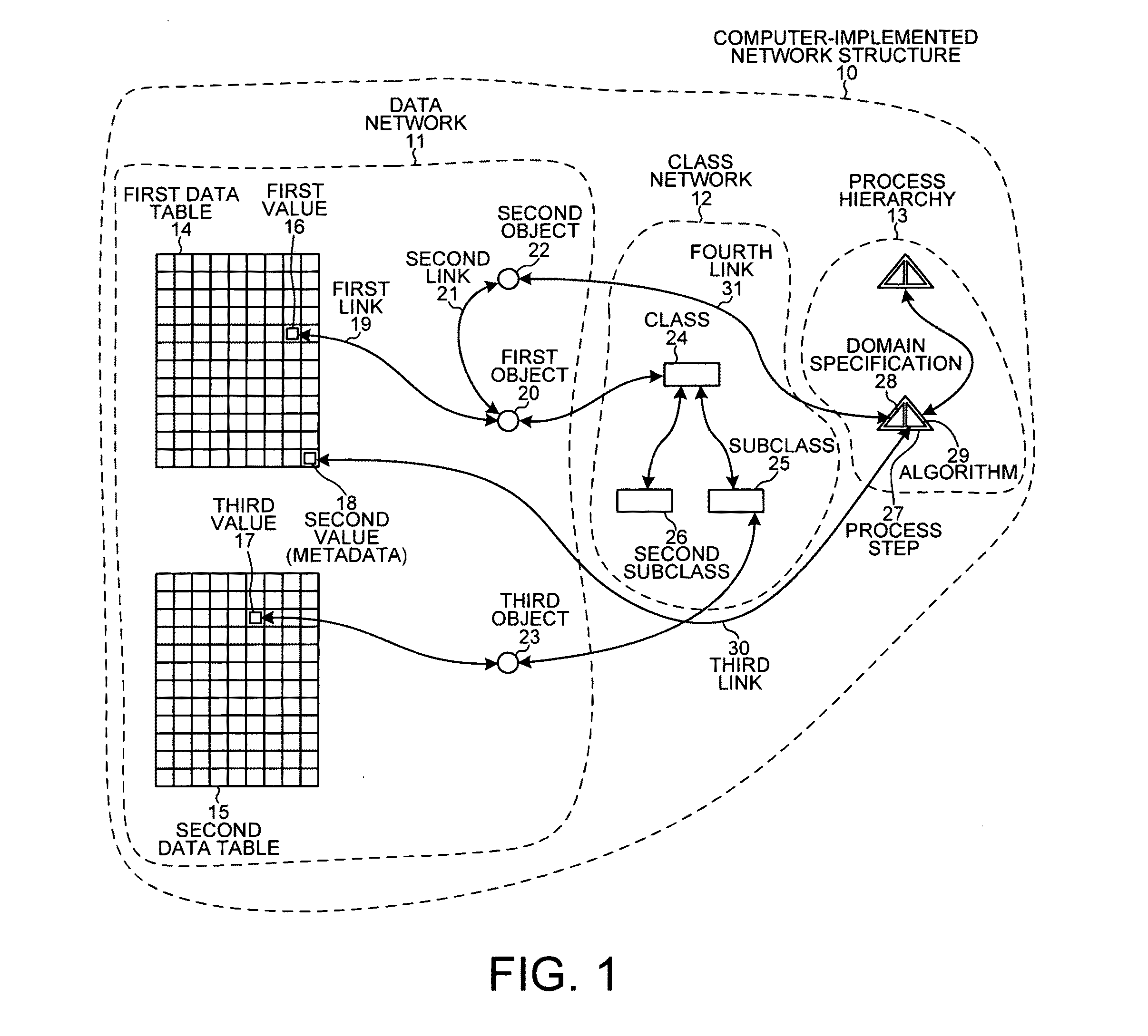
[0061]FIG. 1 is a simplified diagram of a computer-implemented network structure 10 used to locate information of interest from among table data values. The network structure 10 includes a data network 11, a class network 12 and a process hierarchy 13. In the example of FIG. 1, data network 11 includes a first data table 14 and a second data table 15. The table data values in data tables 14-15 are in the form of both numbers and text.

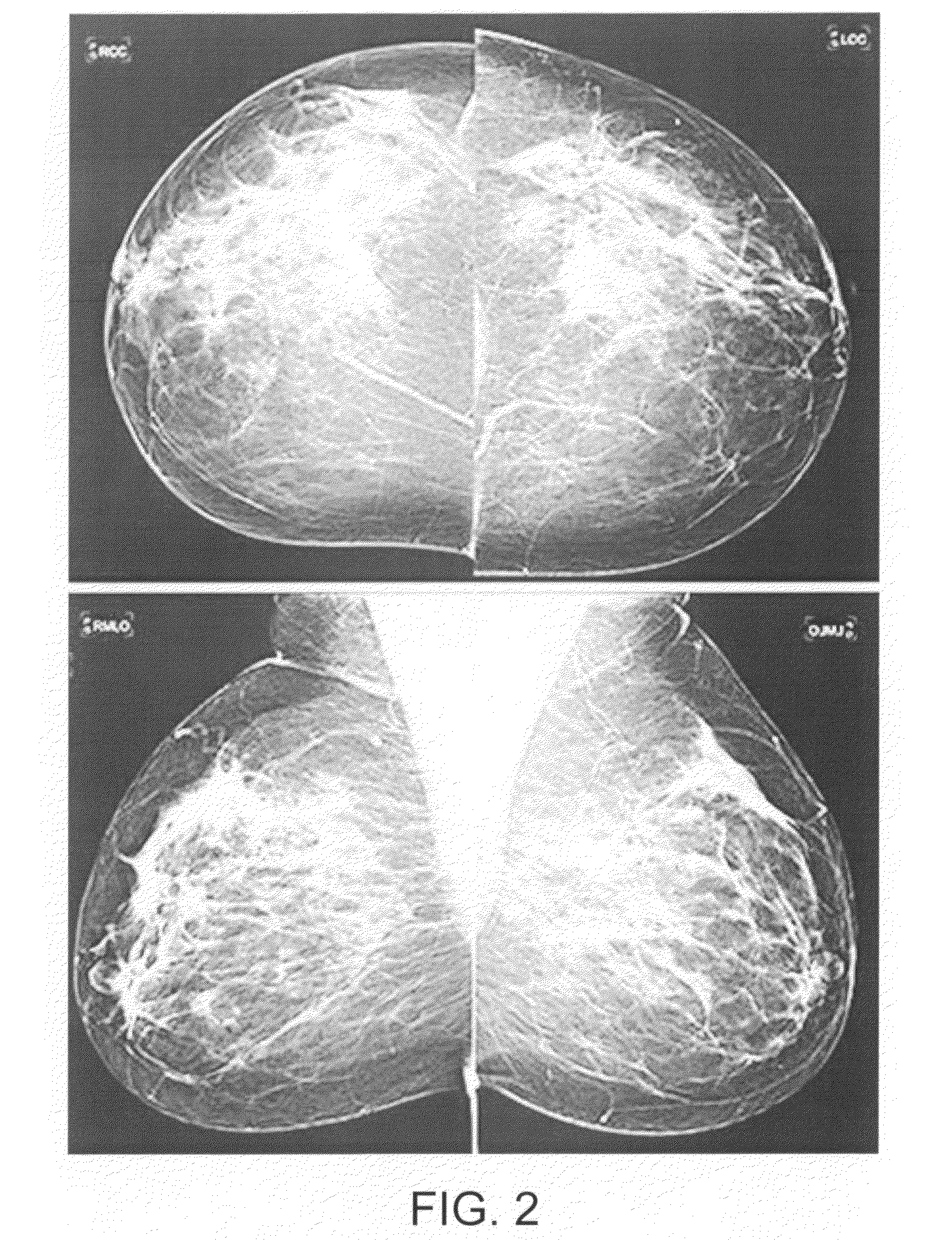
[0062]In one embodiment, some of the table data values describe a medical patient, while other table data values are digital pixel values from a medical image of the patient. The patient is suspected of having breast cancer, and the medical image is a mammogram. Thus, some of the table data values are floating-point values representing the spectral intensity of individual pixels of the digitized mammogram. The o...


PUM


Abstract

An image-based biomarker is generated using image features obtained through object-oriented image analysis of medical images. The values of a first subset of image features are measured and weighted. The weighted values of the image features are summed to calculate the magnitude of a first image-based biomarker. The magnitude of the biomarker for each patient is correlated with a clinical endpoint, such as a survival time, that was observed for the patient whose medical images were analyzed. The correlation is displayed on a graphical user interface as a scatter plot. A second subset of image features is selected that belong to a second image-based biomarker such that the magnitudes of the second image-based biomarker for the patients better correlate with the clinical endpoints observed for those patients. The second biomarker can then be used to predict the clinical endpoint of other patients whose clinical endpoints have not yet been observed.

Description

CROSS REFERENCE TO RELATED APPLICATION[0001]This application is a continuation in part of, and claims priority under 35 U.S.C. §120 from, nonprovisional U.S. patent application Ser. No. 11 / 511,930 entitled “Cognition Integrator and Language,” filed on Aug. 28, 2006, now U.S. Pat. No. 7,873,223. In addition, this application claims the benefit under 35 U.S.C. §119 of provisional application Ser. No. 61 / 459,180, entitled “Content Driven Image Mining to Generate Image-Based Biomarkers”, filed Dec. 8, 2010. Application Ser. No. 11 / 511,930, in turn, claims priority under 35 U.S.C. §120 from PCT application PCT / EP2006 / 061498 filed on Apr. 10, 2006, which in turn is a continuation application of German Application No. DE 102005016290.8 filed on Apr. 8, 2005, in Germany. Application Ser. No. 11 / 511,930 also claims priority under 35 U.S.C. §120 from, nonprovisional U.S. patent application Ser. No. 10 / 687,477 entitled “Extracting Information from Input Data Using a Semantic Cognition Network,...

Claims


Application Information

Patent Timeline
IPC IPC(8): G06T11/20G06K9/00
CPCG06K9/6253G06T7/0012G06T7/0014G06T2207/30068G06T2207/10116G06V10/945G06T2207/30004G06T2207/30061G01N2800/00G06F18/40
Inventor SCHMIDT, GUENTERBINNIG, GERDSCHOENMEYER, RALFSCHAEPE, ARNO
Owner DEFINIENS AG
Why Eureka
Try Eureka