Eureka AIR delivers breakthrough ideas for toughest innovation challenges, trusted by R&D personnel around the world.

Medical image display apparatus, medical image display method and program

Active Publication Date: 2011-08-11
FUJIFILM CORP
View PDF9 Cites 41 Cited by
  • Summary
  • Abstract
  • Description
  • Claims
  • Application Information

AI Technical Summary

Benefits of technology

[0017]The body surface of the examinee is extracted from the three-dimensional medical image, and the medical tomographic image is corrected based on a misalignment between the extracted body surface and the body surface of the model subject. A standard tomographic image corresponding to the corrected medical tomographic image is identified, and then an anatomical tomographic image is identified. Consequently, even if the body surface of the model subject and the body surface of the examinee are not in alignment with each other, an anatomical tomographic image, which is close to the medical tomographic image that is read for diagnosis, can accurately be identified, and an anatomical figure, which is close to the medical tomographic image, can be displayed.

Problems solved by technology

However, it is tedious and time-consuming to use an anatomy atlas, and further, an anatomy atlas does not have all of the sectional figures corresponding to arbitrary tomographic images.
However, the disclosed anatomical figure selecting apparatus fails to display an anatomical image corresponding to an arbitrary tomographic image that the doctor reads for diagnosis.

Method used


Image

  • Medical image display apparatus, medical image display method and program
  • Medical image display apparatus, medical image display method and program
  • Medical image display apparatus, medical image display method and program

Examples


first embodiment

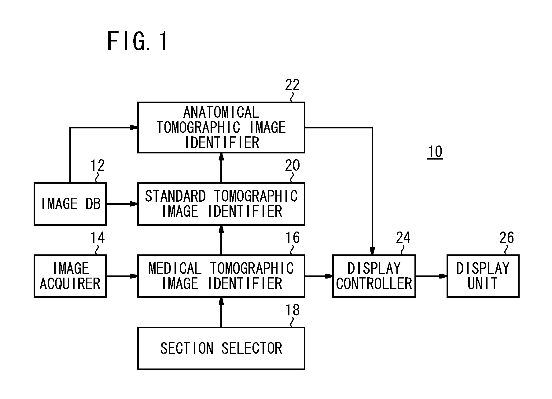
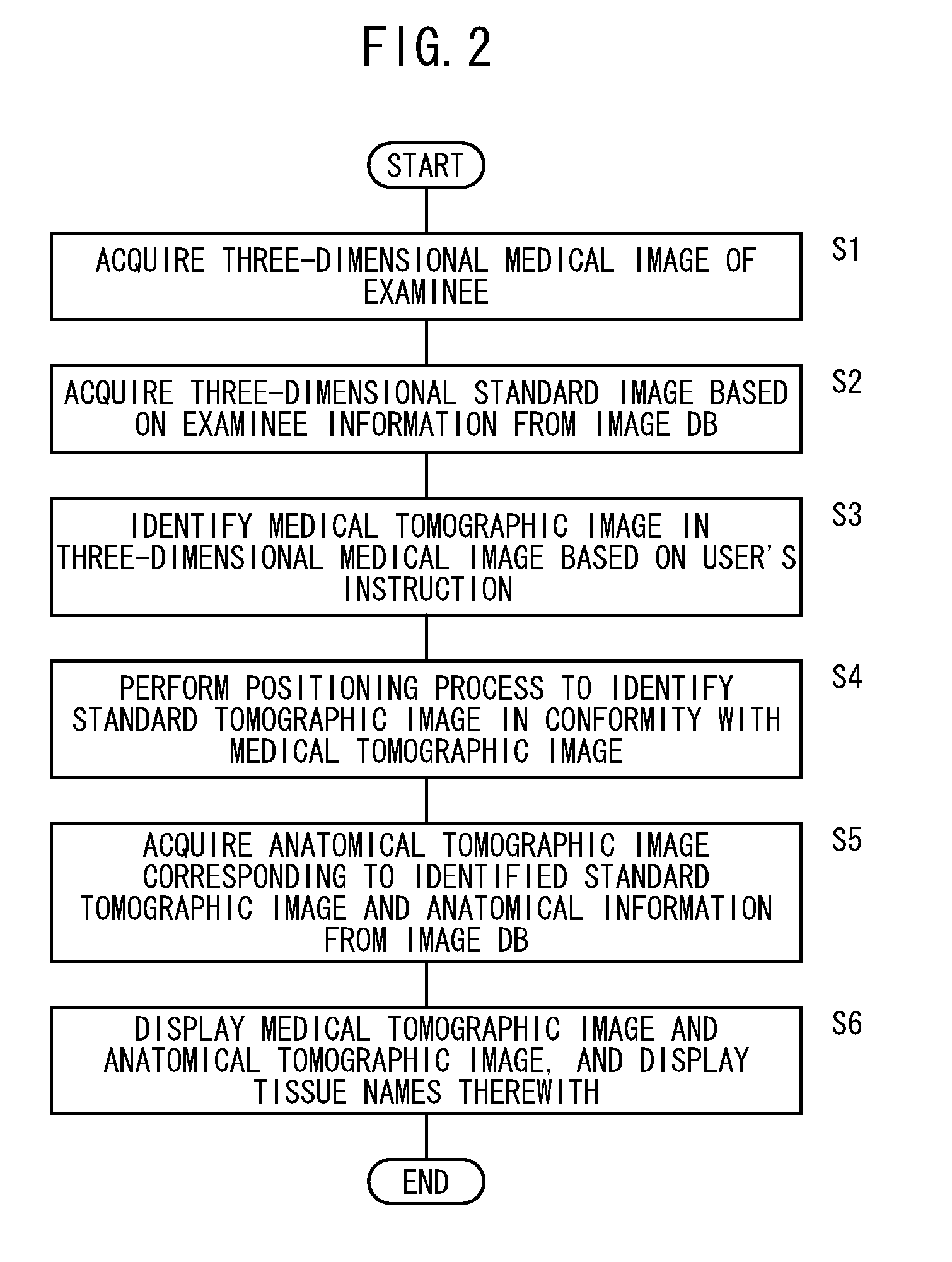
[0030]FIG. 1 is an electric block diagram of a medical image display apparatus 10 according to a first embodiment of the present invention. As shown in FIG. 1, the medical image display apparatus 10 comprises an image database (DB) 12, an image acquirer 14, a medical tomographic image identifier 16, a section selector 18, a standard tomographic image identifier 20, an anatomical tomographic image identifier 22, a display controller 24, and a display unit 26. When an information processor (computer) reads and executes a certain program, the information processor functions as the aforementioned image acquirer 14, the medical tomographic image identifier 16, the standard tomographic image identifier 20, the anatomical tomographic image identifier 22, and the display controller 24.

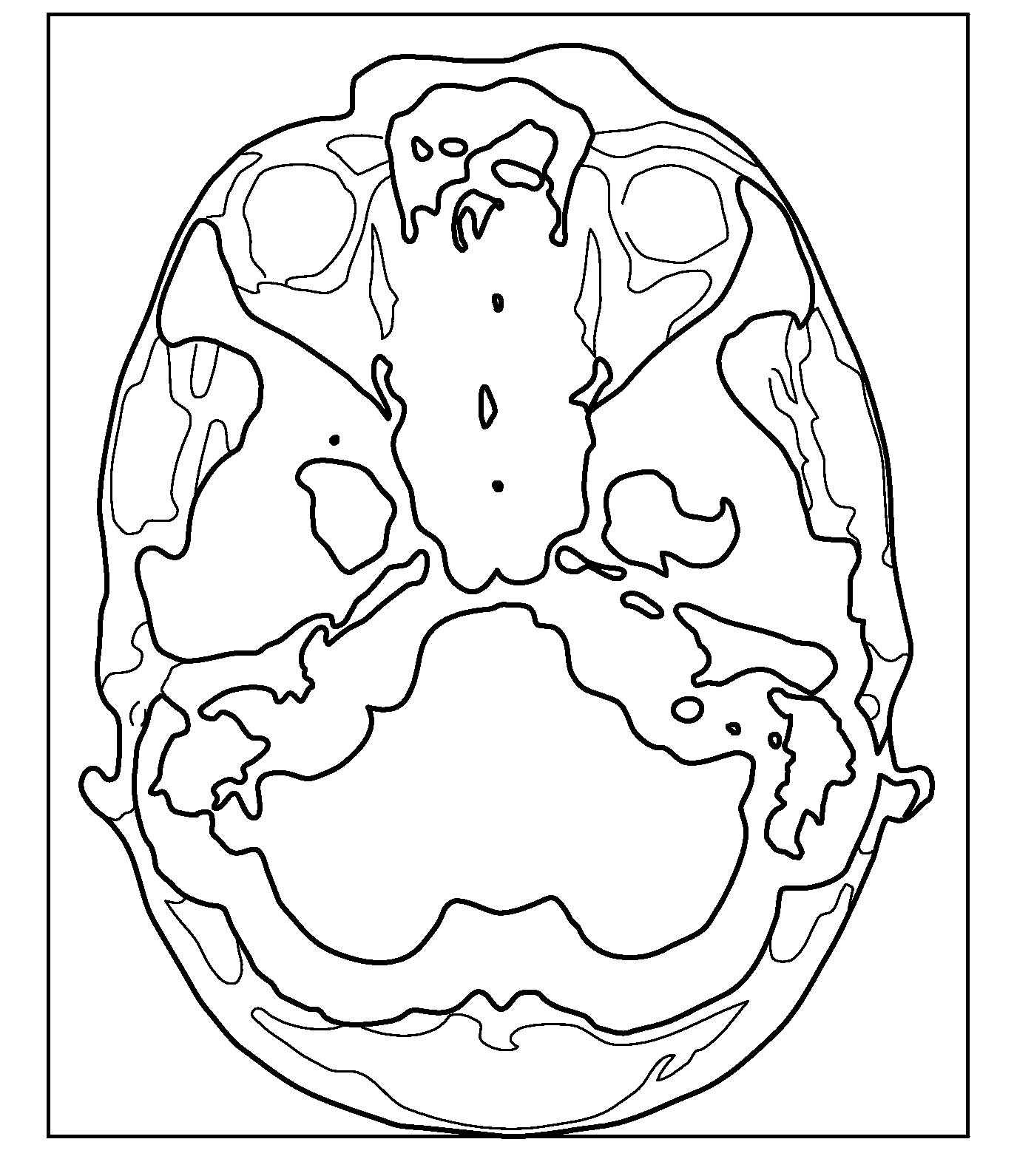
[0031]The image database 12 records therein a three-dimensional standard image of a model subject, a three-dimensional anatomical image corresponding to the three-dimensional standard image, and anatomical inf...

second embodiment

[0049]FIG. 6 is an electric block diagram of a medical image display apparatus 50 according to a second embodiment of the present invention. As shown in FIG. 6, the medical image display apparatus 50 comprises an image database 12, an image acquirer 14, a medical tomographic image identifier 16, a section selector 18, a standard tomographic image identifier 20, an anatomical tomographic image identifier 22, a display controller 24, a display unit 26, a body surface extractor 52, and an image corrector 54. Components of the medical image display apparatus 50, which are identical or equivalent to those of the medical image display apparatus 10 shown in FIG. 1, are denoted by identical reference characters, and such features will not be described in detail below. The image database 12 records therein information representative of a body surface of a model subject, which is represented by a three-dimensional standard image (the region of a model subject in a three-dimensional standard i...


PUM


Abstract

A medical image display apparatus includes an image database for recording a three-dimensional standard image of a model subject, a three-dimensional anatomical image corresponding to the three-dimensional standard image, and anatomical information on living tissue names, an image acquirer for acquiring a three-dimensional medical image of an examinee, a medical tomographic image identifier for identifying a medical tomographic image in the three-dimensional medical image based on a user's instruction, a standard tomographic image identifier for identifying a standard tomographic image in the three-dimensional standard image, which corresponds to the medical tomographic image, an anatomical tomographic image identifier for identifying an anatomical tomographic image in the three-dimensional anatomical image, which corresponds to the standard tomographic image, and a display controller for displaying the medical tomographic image and the anatomical tomographic image on a display unit, while displaying the living tissue names over the anatomical tomographic image based on the anatomical information.

Description

CROSS-REFERENCE TO RELATED APPLICATION[0001]This application is based upon and claims the benefit of priority from Japanese Patent Application No. 2010-024587 filed on Feb. 5, 2010, of which the contents are incorporated herein by reference.BACKGROUND OF THE INVENTION[0002]1. Field of the Invention[0003]The present invention relates to a medical image display apparatus, a medical image display method, and a program for displaying medical tomographic images.[0004]2. Description of the Related Art[0005]Heretofore, in the field of medical images, often it has been customary for a doctor to check names and regions of tissues that are included in displayed images when the doctor reads images for diagnosis. Usually, the doctor checks the images with an anatomy atlas. However, it is tedious and time-consuming to use an anatomy atlas, and further, an anatomy atlas does not have all of the sectional figures corresponding to arbitrary tomographic images. Therefore, an anatomy atlas may not ha...

Claims


Application Information

Patent Timeline
IPC IPC(8): G06K9/00G16H30/40
CPCA61B6/5235G06F19/3437G06F19/321G16H50/50G16H30/40
Inventor WANG, CAIHUAMORIYA, YOSHIYUKI
Owner FUJIFILM CORP
Why Eureka
Eureka Blog
Learn More