Intraspinal Device Deployed Through Percutaneous Approach Into Subarachnoid or Intradural Space of Vertebral Canal to Protect Spinal Cord From External Compression

a technology of intraspinal device and vertebral canal, which is applied in the field of medical devices, can solve the problems of neurological deficit, large process, and segmental sensory loss, and achieve the effect of preventing the development of neurologic deficits

Active Publication Date: 2011-10-27
QURESHI ADNAN IQBAL
View PDF8 Cites 8 Cited by
  • Summary
  • Abstract
  • Description
  • Claims
  • Application Information

AI Technical Summary

Problems solved by technology

The external compression of the spinal cord leads to paraparesis, segmental sensory loss, and sometimes urinary and fecal incontinence.
However, the process is extensive and is associated with complications such as bleeding within the operative sites, or worsening of neurological deficits.

Method used


Image

  • Intraspinal Device Deployed Through Percutaneous Approach Into Subarachnoid or Intradural Space of Vertebral Canal to Protect Spinal Cord From External Compression
  • Intraspinal Device Deployed Through Percutaneous Approach Into Subarachnoid or Intradural Space of Vertebral Canal to Protect Spinal Cord From External Compression
  • Intraspinal Device Deployed Through Percutaneous Approach Into Subarachnoid or Intradural Space of Vertebral Canal to Protect Spinal Cord From External Compression

Examples


Embodiment Construction

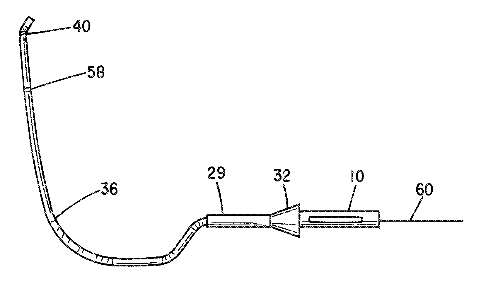
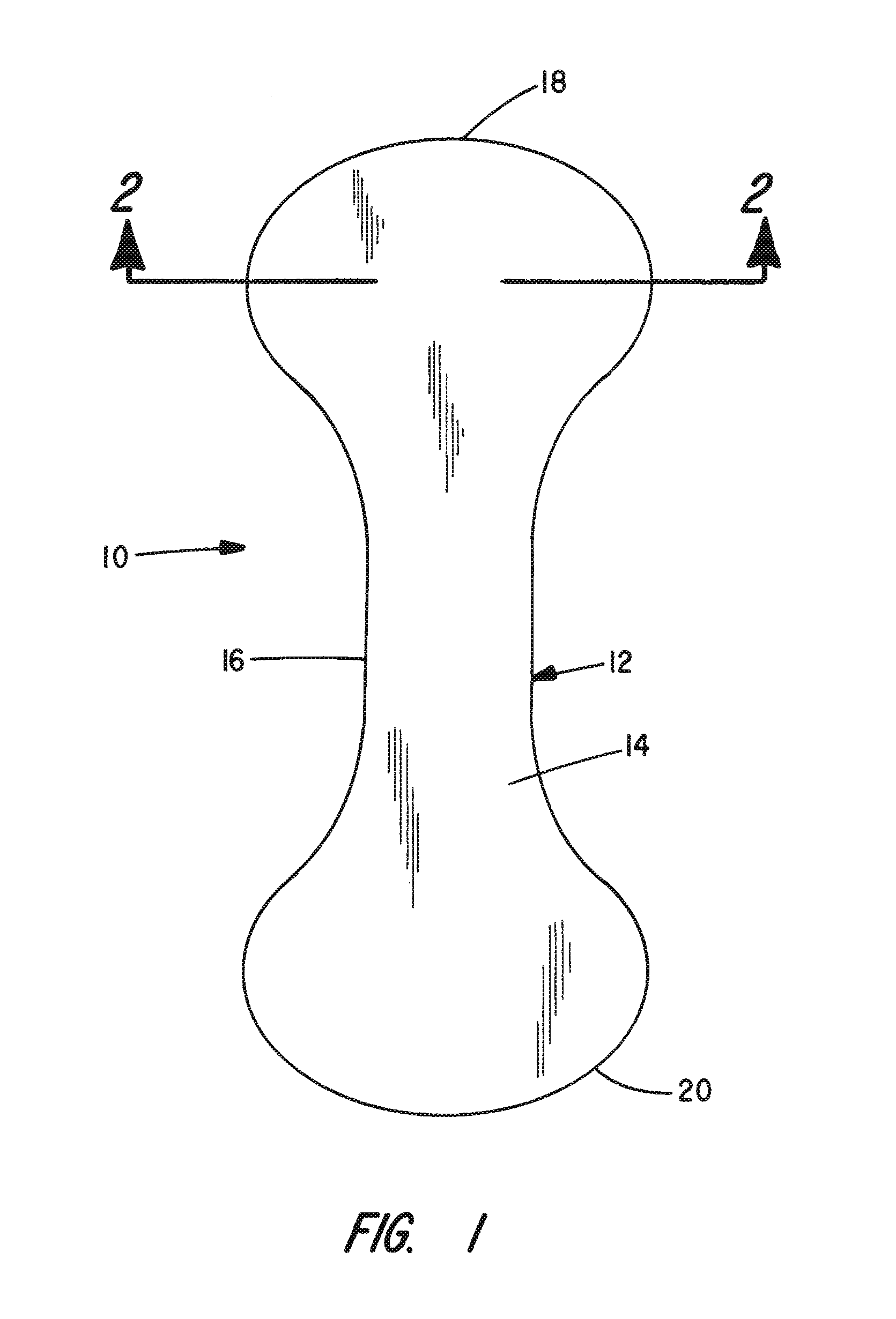
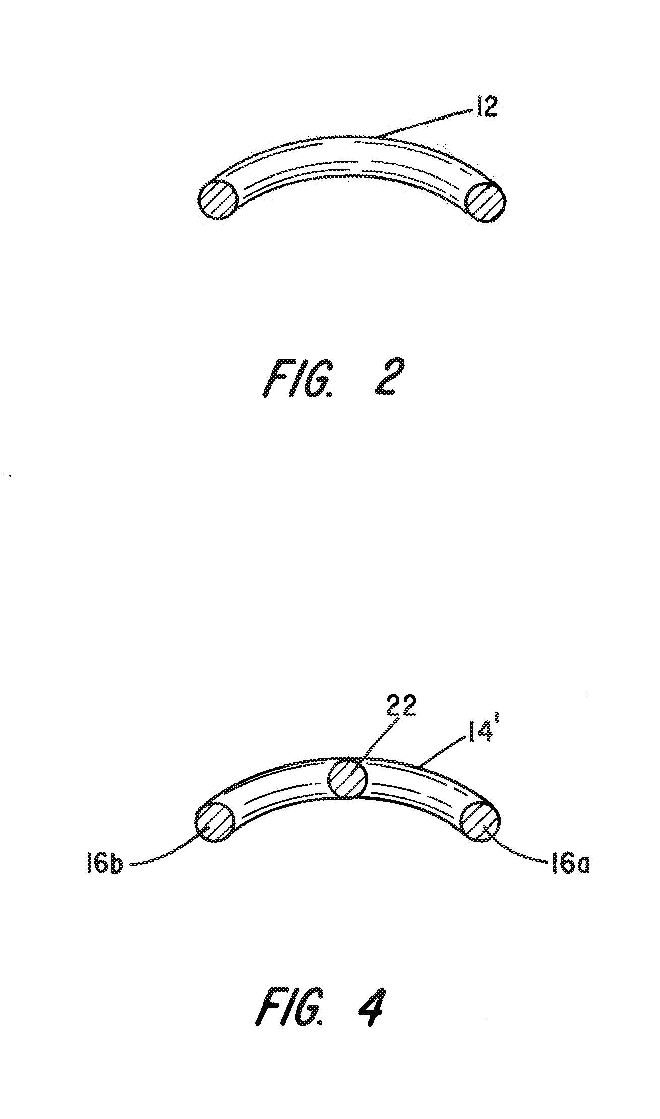
Referring to FIG. 1, the present invention comprises a self-expanding, generally flat, dumbbell-shaped, implantable barrier device 10 which can be introduced in a compressed form through a flexible delivery tube into the subarachnoid or intradural spaces within the vertebral canal under fluoroscopic guidance. The implantable barrier device includes a frame 12 formed from a material exhibiting shape memory properties and consisting of a closed loop of wire where the frame is enclosed within a covering 14 of a selected non-porous membrane material. Without limitation, the frame 12 may be formed from one or more strands of Nitinol wire with plural strands being wound as a cable. The non-porous membrane covering 14 for the frame may be polyurethane, latex or another synthetic material that is somewhat flexible so as to be capable of being readily rolled or folded and which is otherwise body-compatible.

FIG. 1 shows the implantable barrier device 10 in its fully expanded state and the dum...


PUM


Abstract

To shield the spinal cord from an external compression, a barrier device having a self-expanding frame and covered with a non-porous elastomeric sheet is routed through either the subarachnoid or intradural space to the site of the compression through the lumen of a delivery catheter that is percutaneously inserted using an introducer needle. When the distal end of the delivery catheter is proximate the site of the compression, the barrier device is pushed out the distal end of the catheter and allowed to self-expand so as to be interposed between the compression and the spinal cord to prevent impingement.

Description

I. FIELD OF INVENTIONThe present invention relates to a medical device deployed through a minimally invasive procedure to protect the spinal cord from external compression.II. BACKGROUND OF INVENTIONExternal compression of spinal cord through variety of sources including retrograde movement of a vertebral body or disc, a tumor, or a vascular malformation remains major causes of myelopathy. The external compression of the spinal cord leads to paraparesis, segmental sensory loss, and sometimes urinary and fecal incontinence.In humans, the vertebral column is a column usually consisting of 33 vertebrae, the sacrum, intervertebral discs, and the coccyx situated in the dorsal aspect of the torso. The vertebral canal follows the different curves of the spinal column. It is large and triangular in those parts of the column which enjoy the greatest freedom of movement, such as the cervical and lumbar regions and is small and rounded in the thoracic region, where motion is more limited. The ...

Claims


Application Information

Patent Timeline
Patent Type & Authority Applications(United States)
IPC IPC(8): A61F2/44
CPCA61B17/7061A61B17/8858A61F2002/3008A61F2310/00023A61F2002/4435A61F2210/0014A61F2002/30581
Inventor QURESHI, ADNAN IQBAL
Owner QURESHI ADNAN IQBAL
Why Eureka
Try Eureka