

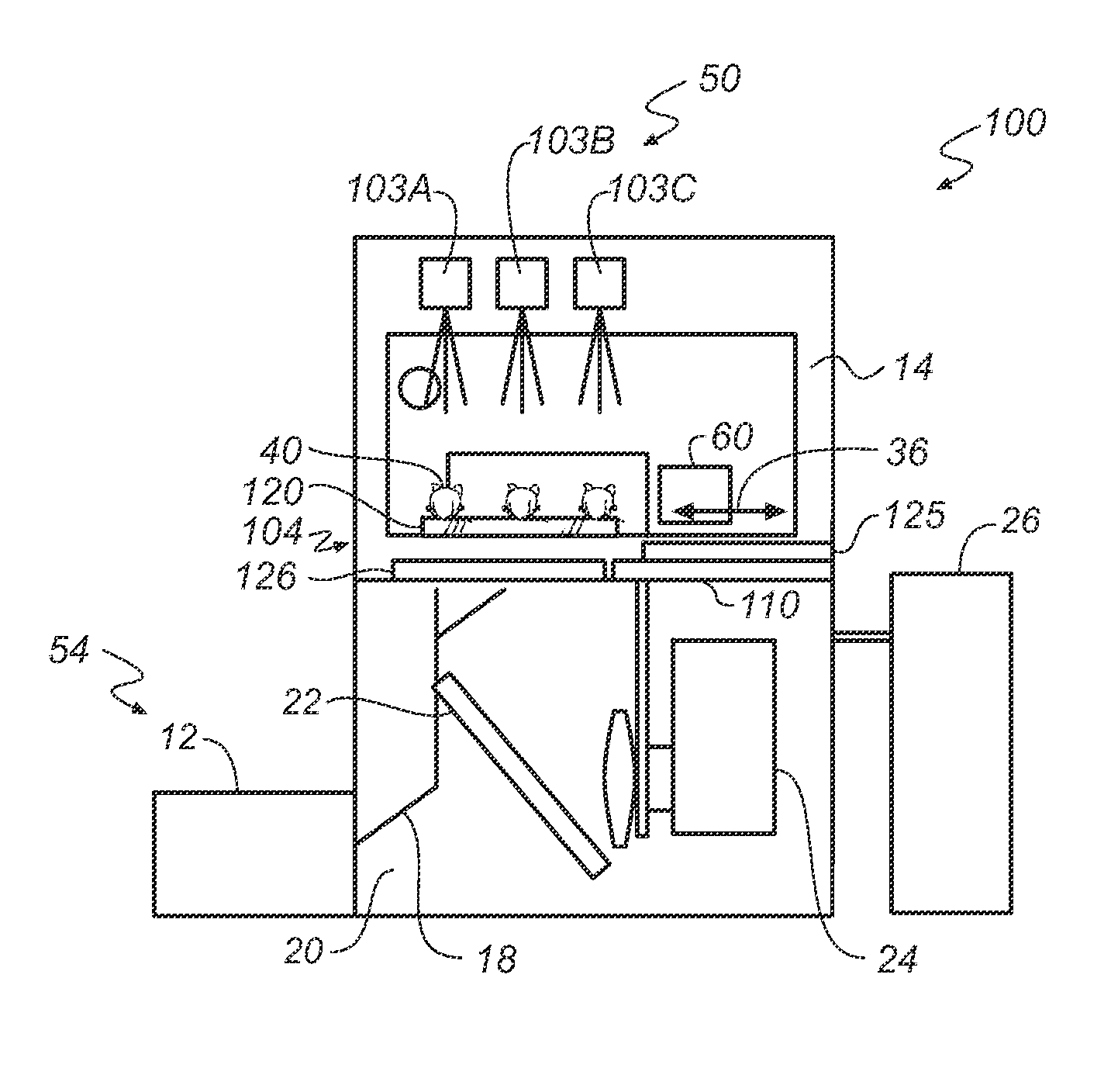
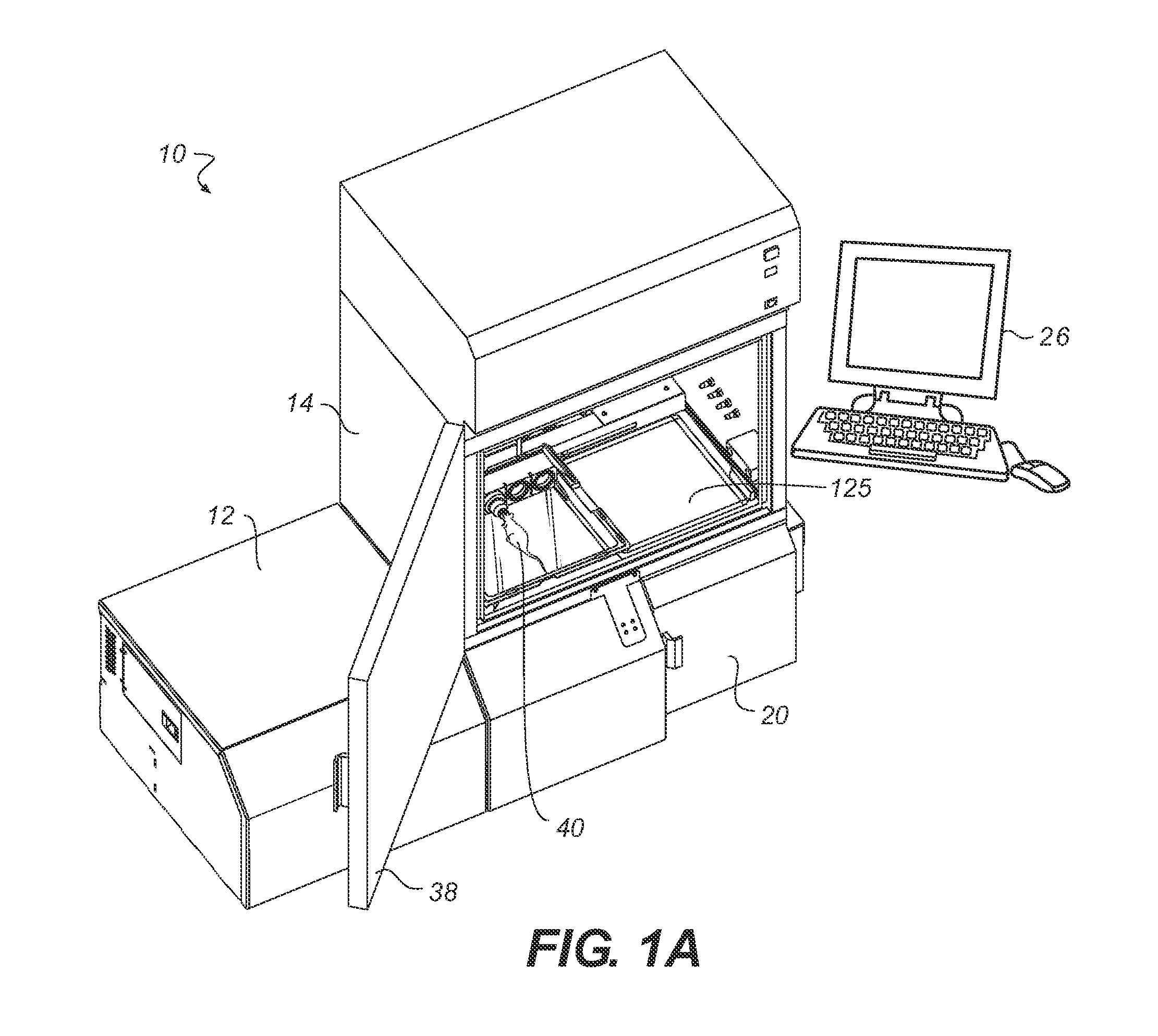
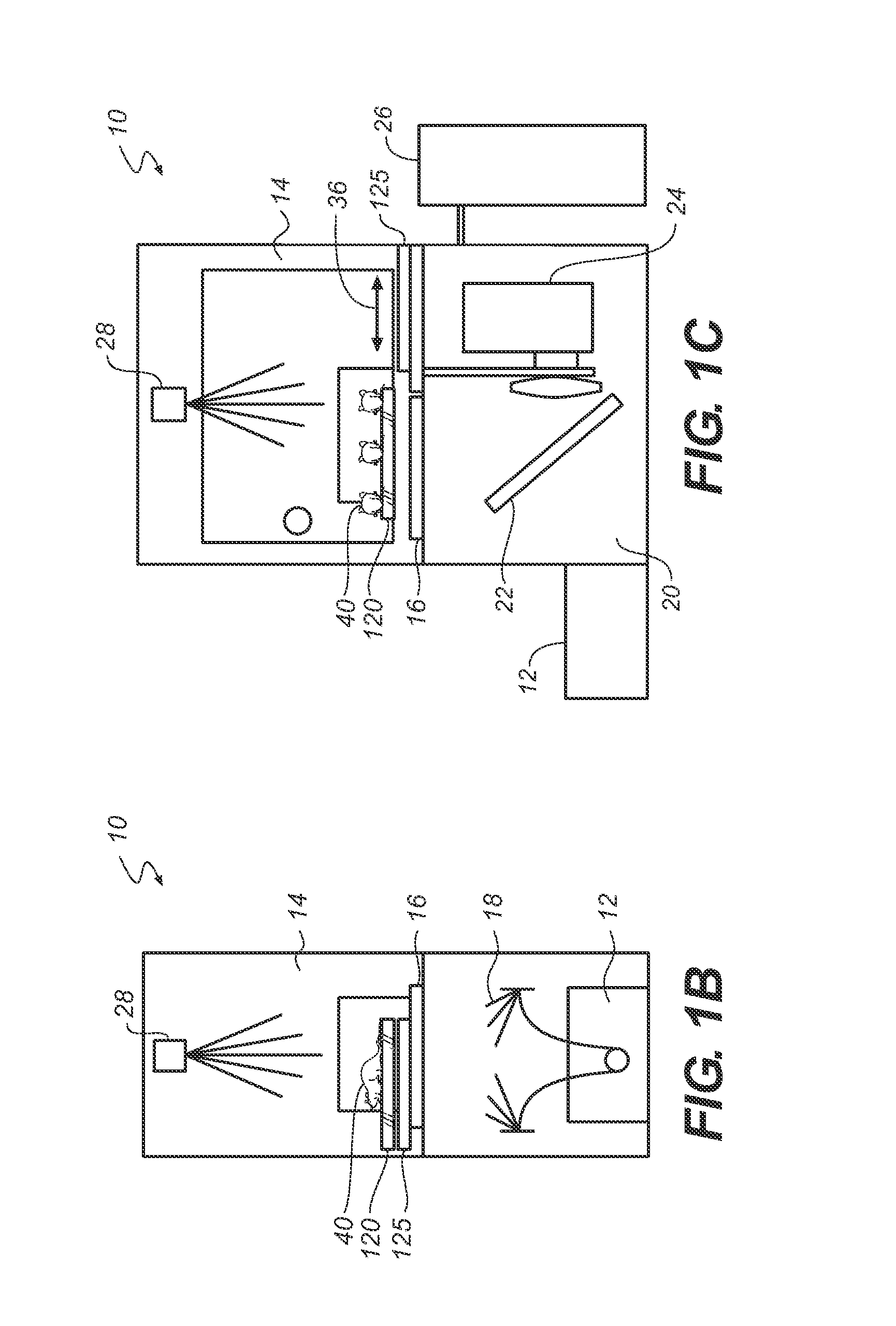
Apparatus and method for multi-modal imaging using multiple x-ray sources
a multi-modal imaging and x-ray source technology, applied in the field of imaging systems, can solve the problems of differential geometric magnification, difficult to correct for geometric magnification in the x-ray image, and greater co-registration error in these systems, so as to reduce the co-registration error among the different modes
- Summary
- Abstract
- Description
- Claims
- Application Information
AI Technical Summary
Benefits of technology
Problems solved by technology
Method used
Image

Examples
Embodiment Construction
[0047]The following is a detailed description of the preferred embodiments of the invention, reference being made to the drawings in which the same reference numerals identify the same elements of structure in each of the several figures. Where they are used, the terms “first”, “second”, and so on, do not necessarily denote any ordinal, sequential, or priority relation, but are simply used to more clearly distinguish one element or set of elements from another, unless specified otherwise.
[0048]Reference is made to U.S. Ser. No. 12 / 196,300 filed Aug. 22, 2008 by Harder et al, entitled APPARATUS AND METHOD FOR MULTI-MODAL IMAGING USING NANOPARTICLE MULTI-MODAL IMAGING PROBES, which published as US 2009 / 0086908.
[0049]Reference is made to U.S. Ser. No. 12 / 354,830 filed Jan. 16, 2009 by Feke et al, entitled APPARATUS AND METHOD FOR MULTI-MODAL IMAGING, which granted as U.S. Pat. No. 8,050,735.
[0050]Reference is made to U.S. Ser. No. 12 / 381,599 filed Mar. 13, 2009 by Feke et al, entitled ...
PUM
 Abstract
Description
Claims
Application Information