Multi-modality contrast and brightfield context rendering for enhanced pathology determination and multi-analyte detection in tissue

a multi-modality contrast and context rendering technology, applied in the field of providing contrast in tissue sections, can solve the problem of unnecessary correlation of differentially labeled features between different tissue sections

US20120200694A1Active Publication Date: 2012-08-09VENTANA MEDICAL SYST INC
13 Cites 60 Cited by

Patent Information

Authority / Receiving Office
US · United States
Current Assignee / Owner
Publication Date
2012-08-09

Smart Images

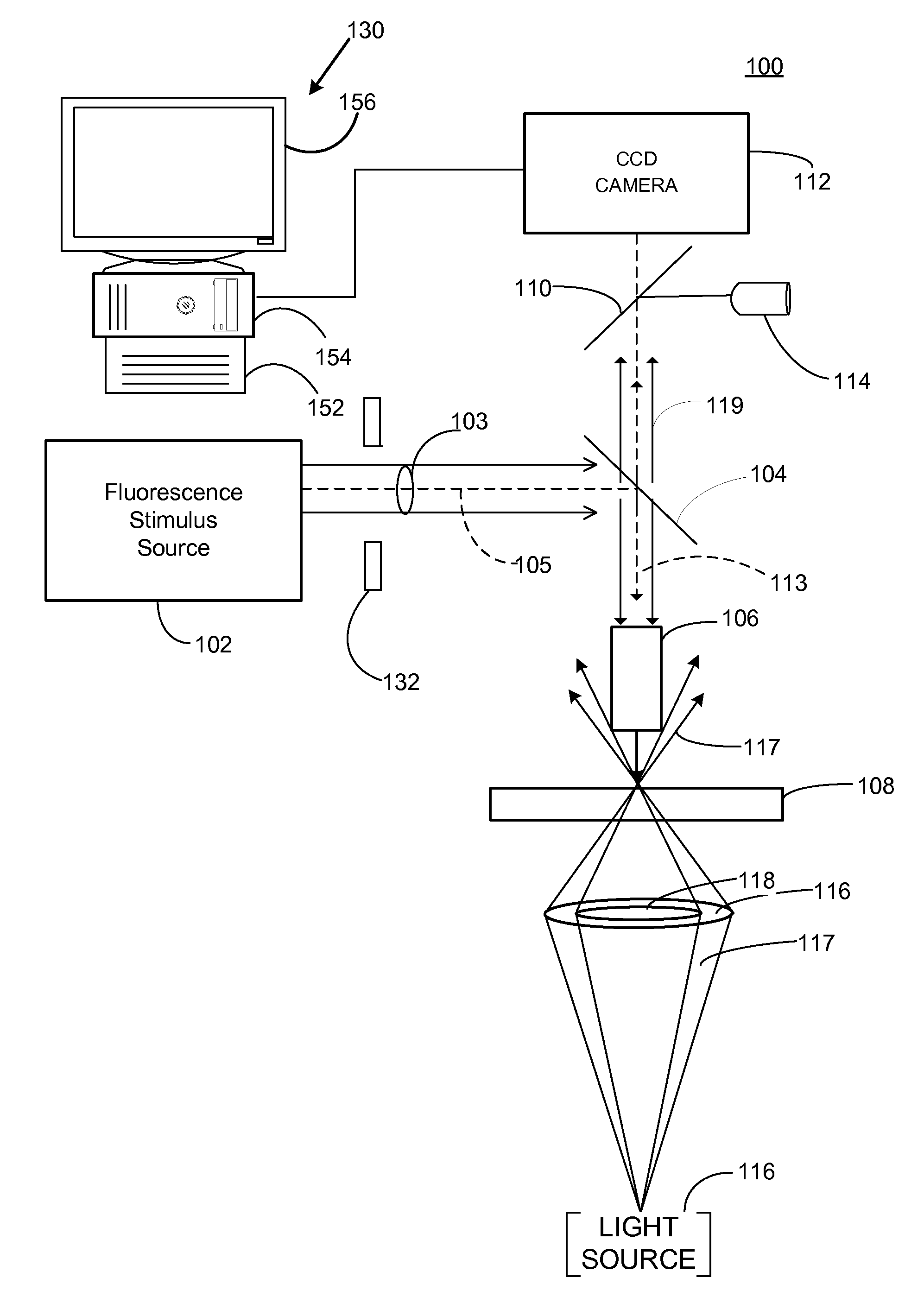
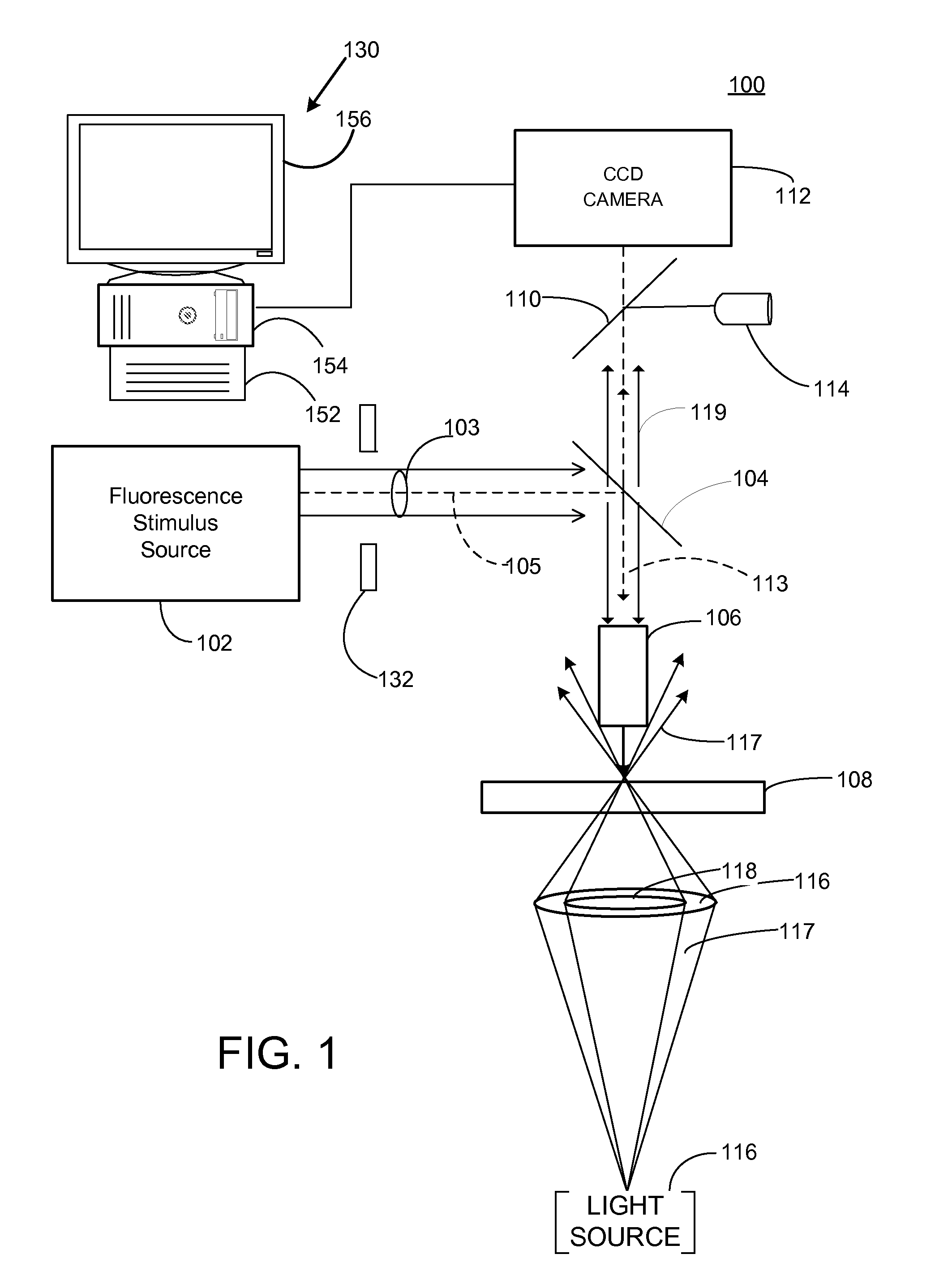
  • Figure 1
    Figure 1
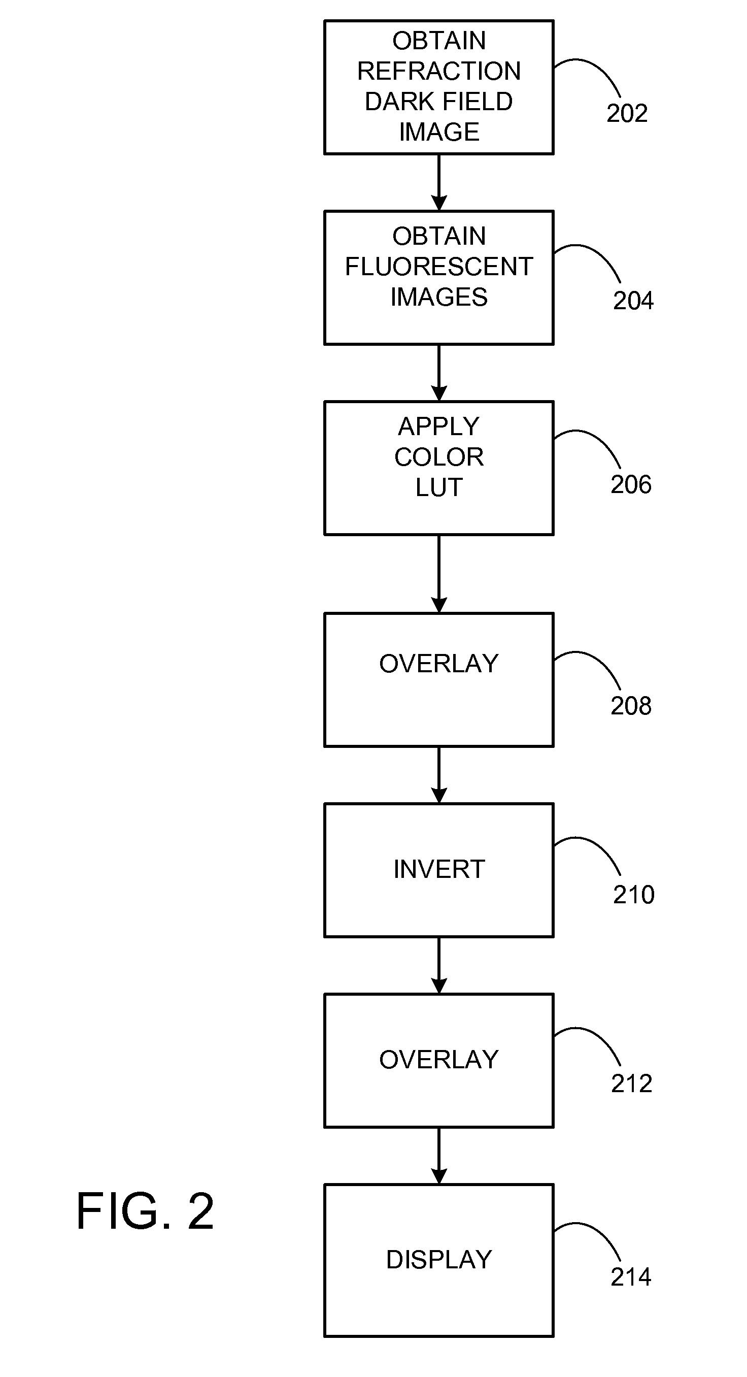
  • Figure 2
    Figure 2
  • Figure 3
    Figure 3
Patent Text Reader

Abstract

Multiple modality contrast can be used to produce images that can be combined and rendered to produce images similar to those produced with wavelength absorbing stains viewed under transmitted white light illumination. Images obtained with other complementary contrast modalities can be presented using engineered color schemes based on classical contrast methods used to reveal the same anatomical structures and histochemistry, thereby providing relevance to medical training and experience. Dark-field contrast images derived from refractive index and fluorescent DAPI counterstain images are combined to produce images similar to those obtained with conventional H&E staining for pathology interpretation. Such multi-modal image data can be streamed for live navigation of histological samples, and can be combined with molecular localizations of genetic DNA probes (FISH), sites of mRNA expression (mRNA-ISH), and immunohistochemical (IHC) probes localized on the same tissue sections, used to evaluate and map tissue sections prepared for imaging mass spectrometry.
Need to check novelty before this filing date? Find Prior Art

Description

CROSS REFERENCE TO RELATED APPLICATIONS

[0001] This application claims the benefit of U.S. Provisional Application No. 61,250,809 filed on Oct. 12, 2009, and U.S. Provisional Application No. 61 / 278,936 filed on Oct. 13, 2009, which applications are incorporated by reference herein in their entirety.FIELD

[0002] The disclosure pertains to methods of providing contrast in tissue sections for pathology determination.BACKGROUND

[0003] Microscopic clinical examination of conventional histological stained tissue sections can be used to evaluate tissue structures and morphological patterns of diagnostic significance. Skilled physicians can view such histological stained tissue sections for diagnosis, and to design and evaluate treatments. The contrast of structures provided by such images using classical stains is familiar, and permits the physician to devote her efforts to interpreting anatomical and morphological tissue section features and anomalies, and not on trying to translate how the sta...

Examples

Embodiment Construction

[0034]As used in this application and in the claims, the singular forms “a,”“an,” and “the” include the plural forms unless the context clearly dictates otherwise. Additionally, the term “includes” means “comprises.”

[0035]The systems, apparatus, and methods described herein should not be construed as limiting in any way. Instead, the present disclosure is directed toward all novel and non-obvious features and aspects of the various disclosed embodiments, alone and in various combinations and sub-combinations with one another. The disclosed systems, methods, and apparatus are not limited to any specific aspect or feature or combinations thereof, nor do the disclosed systems, methods, and apparatus require that any one or more specific advantages be present or problems be solved.

[0036]Although the operations of some of the disclosed methods are described in a particular, sequential order for convenient presentation, it should be understood that this manner of description encompasses r...