Image domain based noise reduction for low dose computed tomography fluoroscopy

a computed tomography and noise reduction technology, applied in tomography, instruments, nuclear engineering, etc., can solve problems such as blurring edges, image noise is increased, and poor image quality

Active Publication Date: 2012-11-22
TOSHIBA MEDICAL SYST CORP
View PDF5 Cites 2 Cited by
  • Summary
  • Abstract
  • Description
  • Claims
  • Application Information

AI Technical Summary

Problems solved by technology

If a lower dose is used, then image noise is increased.
As the tube current is decreased to reduce dose, the image noise increases, resulting in poor image quality.
One method used to reduce image noise is to average the image slices at the same location, but this produces blurring of the edges since there is bound to be movement of the subject, voluntary or involuntary, during the scan.

Method used


Image

  • Image domain based noise reduction for low dose computed tomography fluoroscopy
  • Image domain based noise reduction for low dose computed tomography fluoroscopy
  • Image domain based noise reduction for low dose computed tomography fluoroscopy

Examples


Embodiment Construction

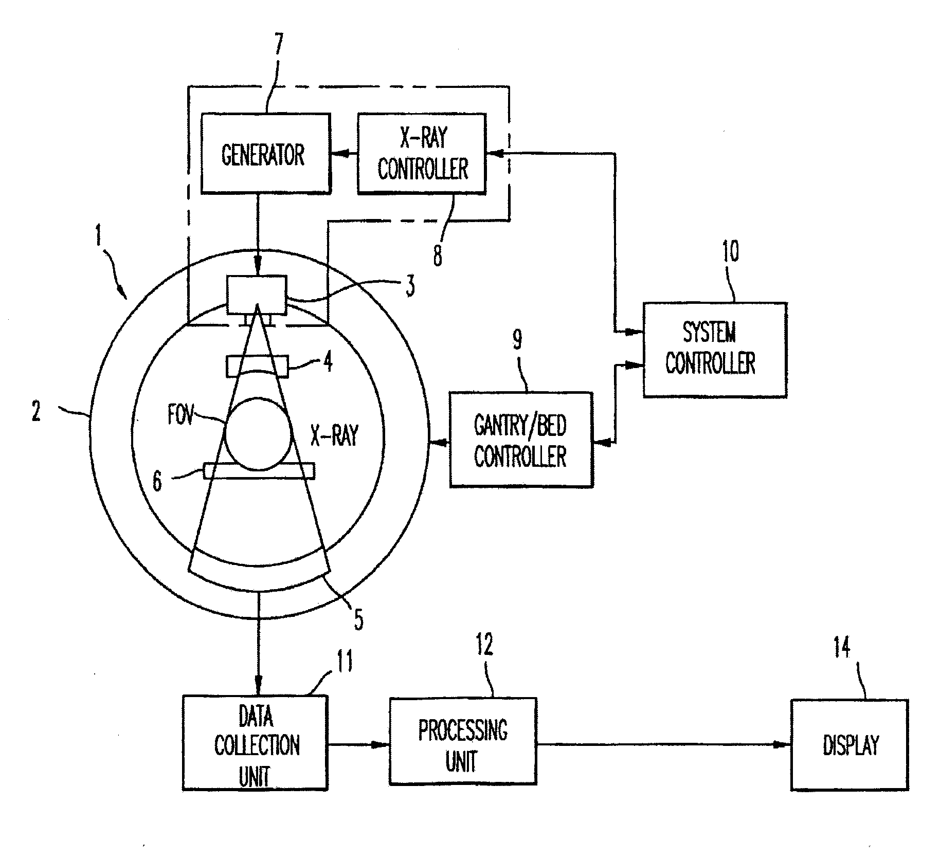
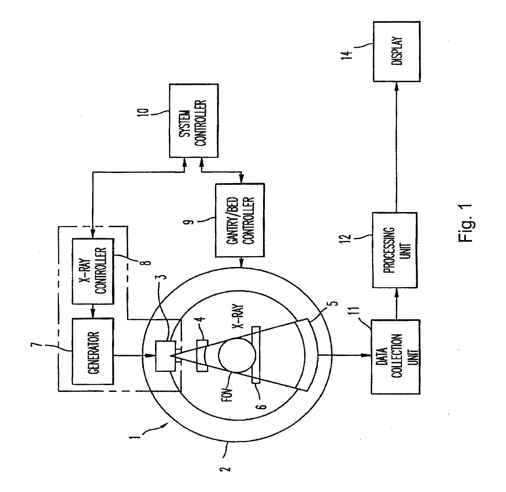
[0021]FIG. 1 shows an x-ray computed tomographic imaging device according to the present invention. The device may be operated as different x-ray doses to carry out different types of scanning, such as CT fluoroscopy. The projection data measurement system constituted by gantry 1 accommodates an x-ray source 3 that generates a cone-beam of x-ray flux approximately cone-shaped, and a two-dimensional array type x-ray detector 5 consisting of a plurality of detector elements arranged in two-dimensional fashion, i.e., a plurality of elements arranged in one dimension stacked in a plurality of rows. X-ray source 3 and two-dimensional array type x-ray detector 5 are installed on a rotating ring 2 in facing opposite sides of a subject, who is laid on a sliding sheet of a bed 6. Two-dimensional array type x-ray detector 5 is mounted on rotating ring 2. Each detector element will correspond with one channel. X-rays from x-ray source 3 are directed on to subject through an x-ray filter 4. X-r...


PUM


Abstract

A method of computed-tomography and a computed-tomography apparatus in which x-ray projection data is acquired at a number of views for a scan of an object. Partial images are created from data for a desired number of said views. Full scan images are created from plural ones of the partial images. Non-overlapping time images are created from the full-scan images. Gradient images are also created. An improved image is created by weighting respective ones of the full scan and non-overlapping time images using the gradient image. The improved image has increased sharpness with reduced noise.

Description

CROSS REFERENCE TO RELATED APPLICATION[0001]This application is a Continuation of and claims the benefit of priority under 35 U.S.C. §120 from U.S. Ser. No. 12 / 539,674, filed Aug. 12, 2009, the contents of which are incorporated herein by reference.BACKGROUND OF THE INVENTION[0002]1. Field of the Invention[0003]The present invention relates to reducing noise in computed tomography (CT) images during CT-fluoroscopy.[0004]2. Discussion of the Background[0005]CT-fluoroscopy involves continuous scanning of a slice or volume of a subject for monitoring in real time, such as monitoring interventions. If a regular dose of x-rays is used, the subject will be exposed to a large x-ray dose. If a lower dose is used, then image noise is increased. In CT, image noise is inversely proportional to the square root of the x-ray tube current. As the tube current is decreased to reduce dose, the image noise increases, resulting in poor image quality. One method used to reduce image noise is to average...

Claims


Application Information

Patent Timeline
Patent Type & Authority Applications(United States)
IPC IPC(8): A61B6/03
CPCG06T11/006Y10S378/901A61B6/03
Inventor MOGHE, SACHINHEIN, ILMAR
Owner TOSHIBA MEDICAL SYST CORP
Why Eureka
Try Eureka