Device and method for illumination of vaginal fornix with ureter location, isolation and protection during hysterectomy procedure

a technology for ureters and vaginal fornixes, which is applied in the field of devices and methods for locating, isolating and protecting ureters during hysterectomy procedures, can solve the problems of inaccurate and time-consuming, inaccurate and time-consuming, and current colpotomizer devices and methods that fail to enable critical visualization of the location of the colpotomizer device, etc., to achieve quick and accurate identification of the incision site

Inactive Publication Date: 2013-05-23
LEXION MEDICAL LLC
View PDF3 Cites 14 Cited by
  • Summary
  • Abstract
  • Description
  • Claims
  • Application Information

AI Technical Summary

Benefits of technology

[0008]The present invention comprises devices and methods that, in certain embodiments, provide a lighted cup, ring or cap that comprises a customizable size and fit, for use in hysterectomy procedures, whereby the ring or cup engages the vaginal fornix and is covered by the vaginal cervical tissue. The lighting allows the surgeon to visualize the location of the lighted cup, ring or cap, thereby quickly and accurately identifying the incision site while providing protection of the associated vasculature and the ureters.

Problems solved by technology

A problem that arises during removal of the uterus is the close anatomical relationship of vascular and ureteral structures that may be injured.
However, current colpotomizer devices and methods fail to enable critical visualization of the location of the colpotomizer device, specifically, the cup ring device.
This failure requires surgeons to awkwardly attempt to locate the device by instrument palpation, a process that is inaccurate and time consuming, requiring repetitive reassessment and introduces the unnecessary hazard of a misguided incision.
During robotic surgeries due to loss of force feedback (haptics), palpation is either severely reduced or not appreciated and can lead to making improperly placed incisions, resulting in unnecessary patient injury.
Such cups often do not provide an optimal fit; the cup selected should just cover the whole cervix.
If, for example, the cup is too large, the ureters will be pulled toward the colpotomy incision, placing the ureters and potentially the uterine artery at risk of damage.
Standard, non-customized sized cups present an unnecessary risk to the patient in cases where the fit is not optimal.

Method used


Image

  • Device and method for illumination of vaginal fornix with ureter location, isolation and protection during hysterectomy procedure
  • Device and method for illumination of vaginal fornix with ureter location, isolation and protection during hysterectomy procedure
  • Device and method for illumination of vaginal fornix with ureter location, isolation and protection during hysterectomy procedure

Examples


Embodiment Construction

[0021]While the invention is amenable to various modifications and alternative forms, specifics thereof are shown by way of example in the drawings and described in detail herein. It should be understood, however, that the intention is not to limit the invention to the particular embodiments described. On the contrary, the intention is to cover all modifications, equivalents, and alternatives falling within the spirit and scope of the invention.

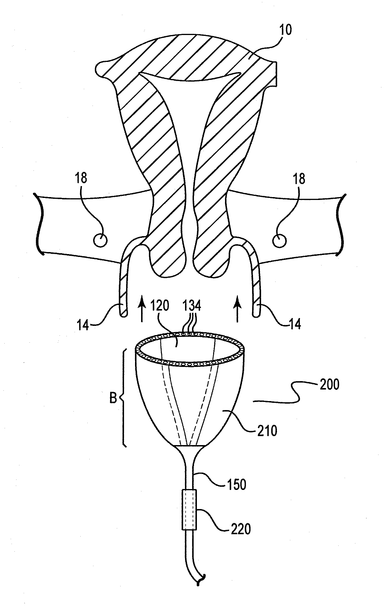
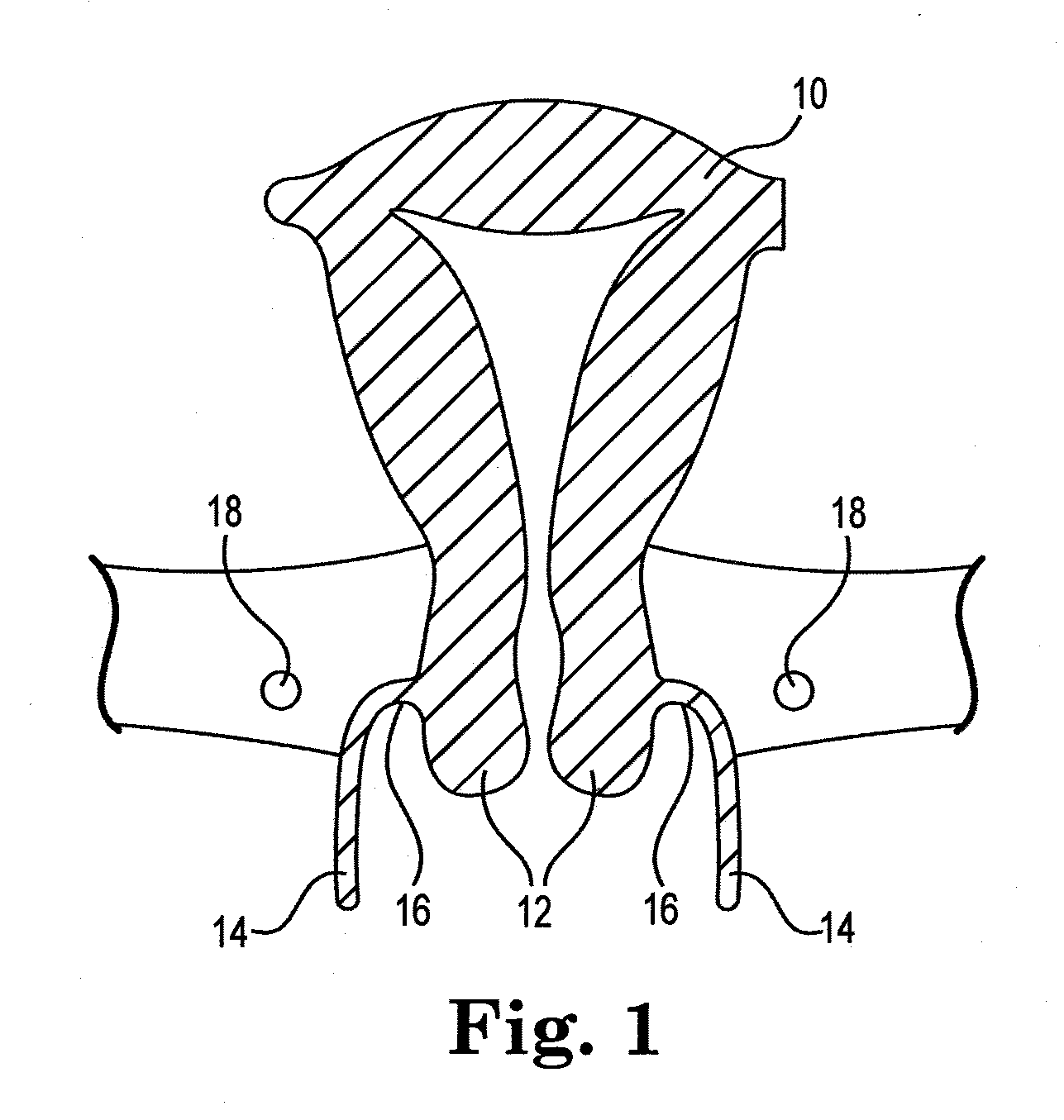
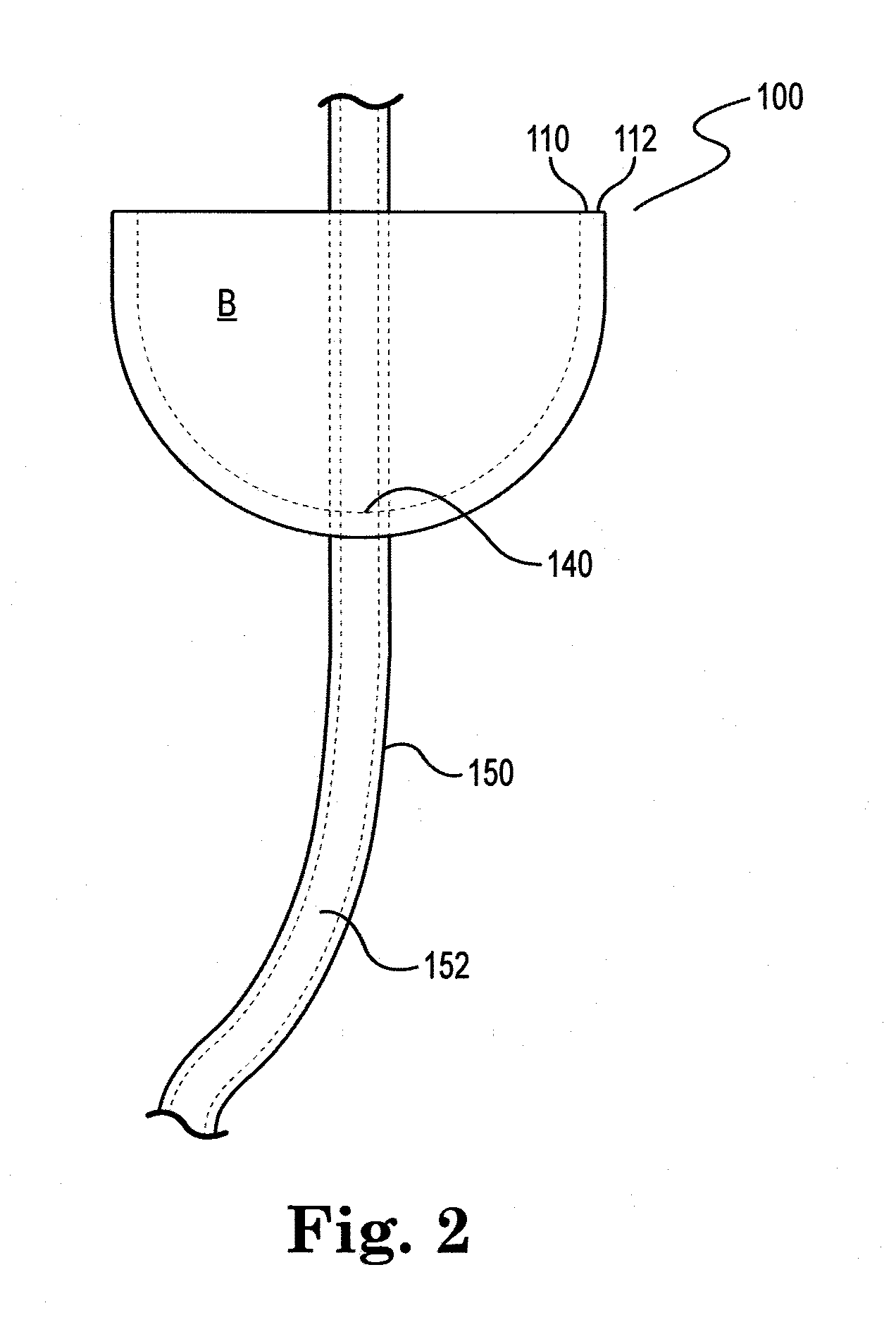
[0022]The present invention comprises devices and methods that overcome persistent difficulties in hysterectomy procedures. In various embodiments, a lighted cervical cup comprising lighting elements disposed around the outer lip of the body of the cervical cup is provided. As defined herein “cup” also comprises a cap or ring which are lighted. The lighting elements are sufficiently bright and of appropriate wavelength to allow penetration of the vaginal cervical tissue covering the lighted cup. This allows visualization of the vaginal cervic...


PUM


Abstract

The present invention comprises devices and methods that, in certain embodiments, provide a lighted cup, ring or cap that comprises a customizable size and fit, for use in hysterectomy procedures, whereby the ring or cup engages the vaginal fornix and is covered by the vaginal cervical tissue. The lighting allows the surgeon to visualize the location of the lighted cup, ring or cap, thereby quickly and accurately identifying the incision site while providing protection of the associated vasculature and the ureters.

Description

CROSS-REFERENCE TO RELATED APPLICATIONS[0001]The present application claims priority to provisional application No. 61 / 560,899, filed on Nov. 17, 2011 and entitled “Lighting Apparatus and Method for Ureter Location, Isolation and Protection During Hysterectomy Procedure”, the entire contents of which are incorporated herein by reference.BACKGROUND OF THE INVENTION[0002]1. Field of the Invention[0003]The invention relates to devices and methods for locating, isolating and protecting ureters during hysterectomy procedures, in certain embodiments providing an illuminating cup or ring or cap over which vaginal cervical tissue is positioned.[0004]2. Description of the Related Art[0005]Known methods of removal of the uterus consist of minimally invasive methods such as laparoscopy and robotic laparoscopic methods. See, e.g., U.S. Pat. No. 5,520,698, describing the primary approaches. A problem that arises during removal of the uterus is the close anatomical relationship of vascular and ur...

Claims


Application Information

Patent Timeline
Patent Type & Authority Applications(United States)
IPC IPC(8): A61B1/07A61B1/06
CPCA61B1/06A61B19/5202A61B2017/00115A61B2017/00867A61B2019/4857A61B2019/465A61B2019/5206A61B2019/521A61M2025/0166A61B17/4241A61B2019/5204A61B90/30A61B2090/309A61B2090/065A61B2090/0811A61B2090/304A61B2090/306A61B2090/3945
Inventor WILLIAMS, STEVENOTT, DOUGLAS
Owner LEXION MEDICAL LLC
Why Eureka
Try Eureka